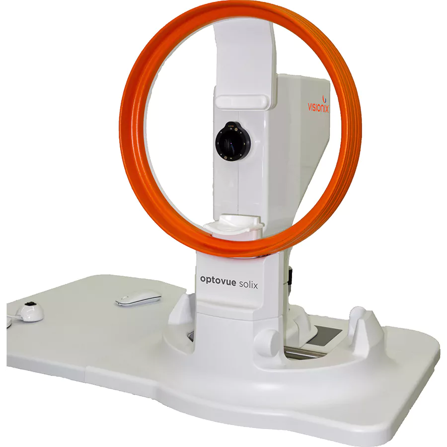
Next generation imaging from cornea to choroid.
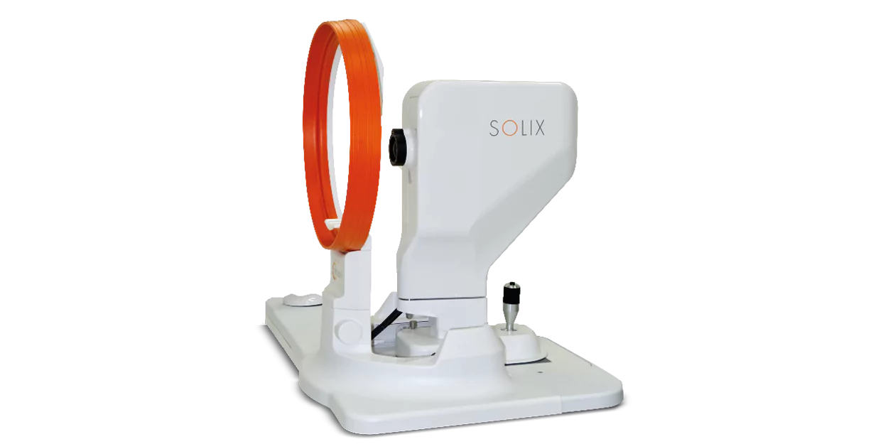
Optovue Solix Essential is a technology built upon a proven foundation of high-speed Spectral Domain OCT. The Optovue Solix Essential offers state-of-the-art imaging from the cornea to the choroid with exclusive technology that will change your approach to disease diagnosis and management.
New advanced High-density (HD) retina scan patterns for maximum resolution and post-processing alignment. Tracked High-density scans with SSADA & MCT with vessel-to-vessel post-processing alignment that produces a superior platform for change as it minimizes scan location and movement effects during acquisition and allows for precise registration.
New advanced scans and glaucoma analytics take glaucoma scanning to the next level, incorporating Dual Track, SSADA, MCT, and AI segmentation with repeatability and reproducibility 2 times better than before to make a precision glaucoma system.
Comprehensive anterior evaluation of pathologies such as keratoconus and dry eye symptoms utilizing pachymetry and epithelial thickness mapping, and 3D EnFace imaging.
iWellness capabilities have become part of a standard of care for patients suspected of retinal pathologies and/or glaucoma. The Solix AngioWellness scan enables a comprehensive assessment of your vasculature for diabetic patients and nerve fiber loss (glaucoma) suspects by combining structural information on retinal and ganglion cell thickness with objective metrics on retinal vasculature. Utilizing FAZ Analytics to uncover early indicators of vascular changes associated with diabetic patients.

SOLIX ESSENTIAL TECHNICAL SPECIFICATIONS
OCT Imaging | Retina
OCT-A Imaging
OCT Imaging | Anterior Segment
Electrical and Physical Specifications
Computer/Networking Specifications

Screening for refractive surgery
Raise awareness on the importance of Epithelial Thickness Mapping (ETM) in refractive surgery screening.

Screening for refractive surgery
Raise awareness on the importance of Epithelial Thickness Mapping (ETM) in refractive surgery screening.

