


| Feature | Value |
|---|---|
| Magnifications | 2x/3x/5x |
| Convergent version | • |
| Height adjustment: | • |
| Slit width | 0 – 14 mm Continually |
| Slit length | 0 – 14 mm Continually |
| Slit apertures | 14; 10; 6; 4; 3; 1; 0,2mm |
| Tyndall point | Ø 0,2mm |
| Slit rotation | ± 90° Continually on TABO scheme |
| Working distance | 100mm |
| Average viewing height | 375mm |
| Light | LED |
| Max light intensity | 300 000 Lux |
| Longitudinal (In/Out) | 99mm |
| Lateral (Left/Right) | 118mm |
| Vertical (Up/Down) | 30mm |
| Chin-rest height | 76mm |
| Stereo angle | 6° |
| Eyepiece | 10x |
| Total magnification/field of view (in mm) for 2 magnifications | 10x/27; 16x/16 |
| Total magnification/field of view (in mm) for 3 magnifications | 10x/27; 16x/16; 25x/11 |
| Total magnification/field of view (in mm) for 5 magnifications | 6x/43; 10x/27; 16x/16; 24x/11; 40x/7 |
| Pupillary adjustement | 52 – 90mm (2 magnifications) 55 – 75mm (3 or 5 magnifications) |
| Diopter ajustment | +/- 6D |
| Blue (fluorescence) | • |
| Green (red-free) | • |
| Grey (anti heat) | • |
| Power supply | Provided |
| Power supply for the slit lamp | 3,4V, 700mA |
| Power supply for the fixation point | 5v |
| Input voltage | 110V / 220V AC; 60/50Hz |
| Fusible | NA |
| Width | 380mm |
| Depth | 530mm |
| Height | 780mm |
| Weight | Packed: 26Kg (2 magnifications), 28Kg (3 or 5 magnifications) |
| MDD, CE | • |
| Safety class | I |
| Parts applied | Type B |
| Tonometer | 8480-5010-00 |
| Tonometer support plate | Provided with the tonometer |

Screening for refractive surgery
Raise awareness on the importance of Epithelial Thickness Mapping (ETM) in refractive surgery screening.

Retina protocol
SOLIX delivers pristine images of retinal structures with unprecedented views of the vitreous and choroid, that enable confident diagnosis and management of retinal pathologies.
Protocolo Glaucoma
The SOLIX glaucoma package delivers in-depth analysis of the optic nerve head structure and vasculature. Optovue-exclusive scans bring additional insights that aid in clinical decision making.
Protocole Glaucome
The SOLIX glaucoma package delivers in-depth analysis of the optic nerve head structure and vasculature. Optovue-exclusive scans bring additional insights that aid in clinical decision making.
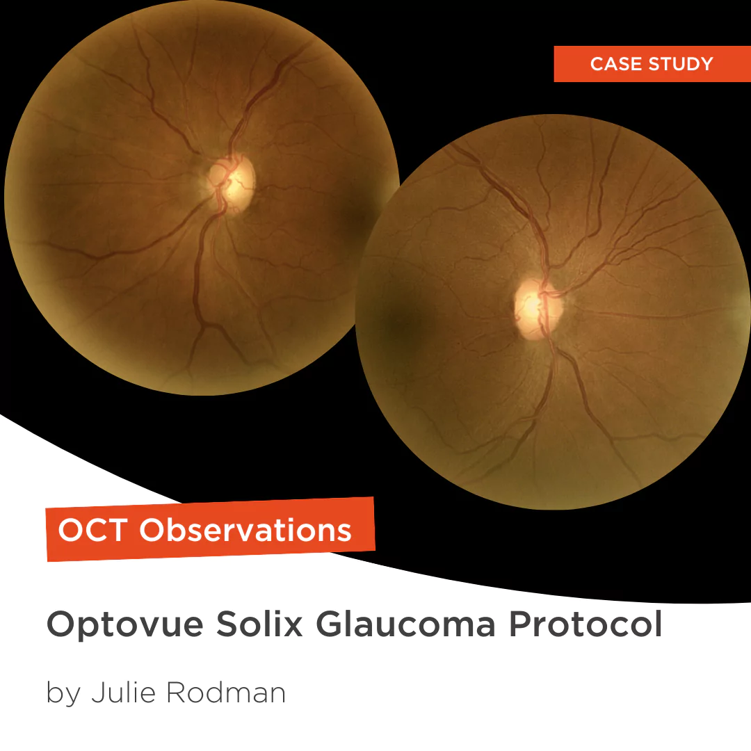
Glaucoma protocol
The SOLIX glaucoma package delivers in-depth analysis of the optic nerve head structure and vasculature. Optovue-exclusive scans bring additional insights that aid in clinical decision making.

We are big fans of Briot
Dr. Gray Sass, OD Wildwood Eyecare
Screening for refractive surgery
Raise awareness on the importance of Epithelial Thickness Mapping (ETM) in refractive surgery screening.
Retina protocol
SOLIX delivers pristine images of retinal structures with unprecedented views of the vitreous and choroid, that enable confident diagnosis and management of retinal pathologies.
Protocolo Glaucoma
The SOLIX glaucoma package delivers in-depth analysis of the optic nerve head structure and vasculature. Optovue-exclusive scans bring additional insights that aid in clinical decision making.
Protocole Glaucome
The SOLIX glaucoma package delivers in-depth analysis of the optic nerve head structure and vasculature. Optovue-exclusive scans bring additional insights that aid in clinical decision making.
Glaucoma protocol
The SOLIX glaucoma package delivers in-depth analysis of the optic nerve head structure and vasculature. Optovue-exclusive scans bring additional insights that aid in clinical decision making.
We are big fans of Briot
Dr. Gray Sass, OD Wildwood Eyecare

My office has had a Briot edger for over 10 years
Dr. Albert Morier Consumer Optical

”Implementing the Briot® Attitude 2 has been effortless. It saves me time so I don’t need to spend extra hours in the office.“
Dr. Douglas King Owner, Family Eye Care Optometry Center

Dr. Paul Karpecki Kentucky Eye Institute
Dr. Paul Karpecki Kentucky Eye Institute

Dr. John Gelles is the director of the specialty contact lens division at The Cornea and Laser Eye Institute – Hersh Vision Group and The CLEI Center for Keratoconus.
Dr. John Gelles is the director of the specialty contact lens division at The Cornea and Laser Eye Institute – Hersh Vision Group and The CLEI Center for Keratoconus.

“I can safely and efficiently practice while also giving the patient confidence they are receiving top notch and modern care.”
Jordan Jones, O.D. Director of Eyecare, Wear Eyewear