


SOLIX delivers pristine images of retinal structures with unprecedented views of the vitreous and choroid, enabling confident diagnosis and management of retinal pathologies – even in highly myopic patients. A precision OCTA scan that generates both RDB metrics and OCTA vessel density for comprehensive retinal analysis, which optimizes efficiency and quickly provides the clinical data your practice demands.

SOLIX delivers multiple tools for a new generation of disease management that improves throughput and enables superior patient care: anterior and posterior OCT, OCT angiography, fundus photography, external color photography, evaluation of the meibomian glands.

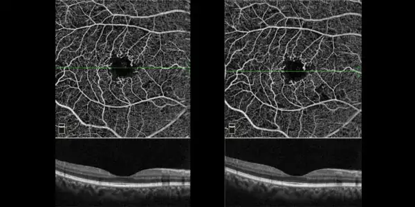
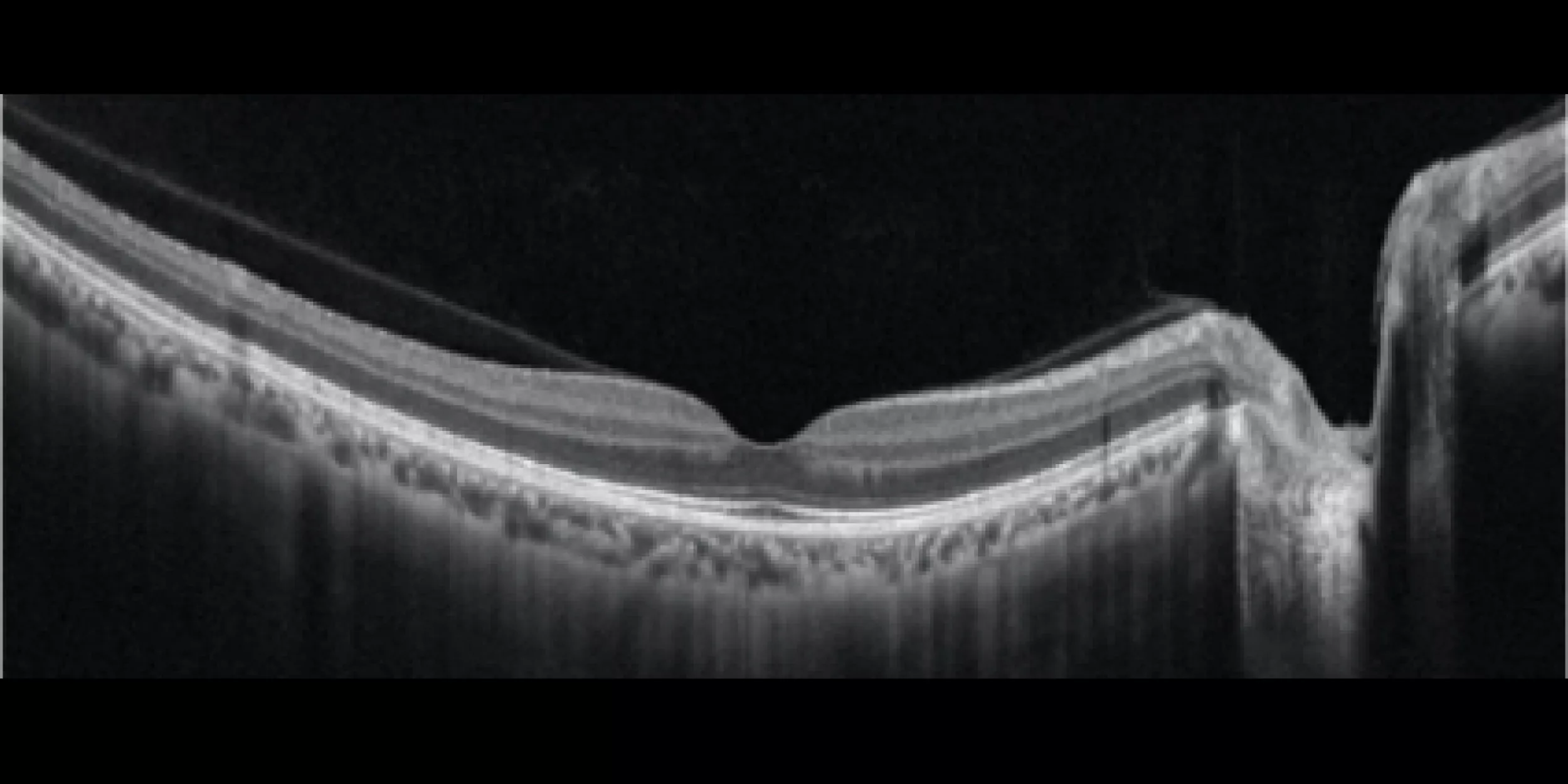
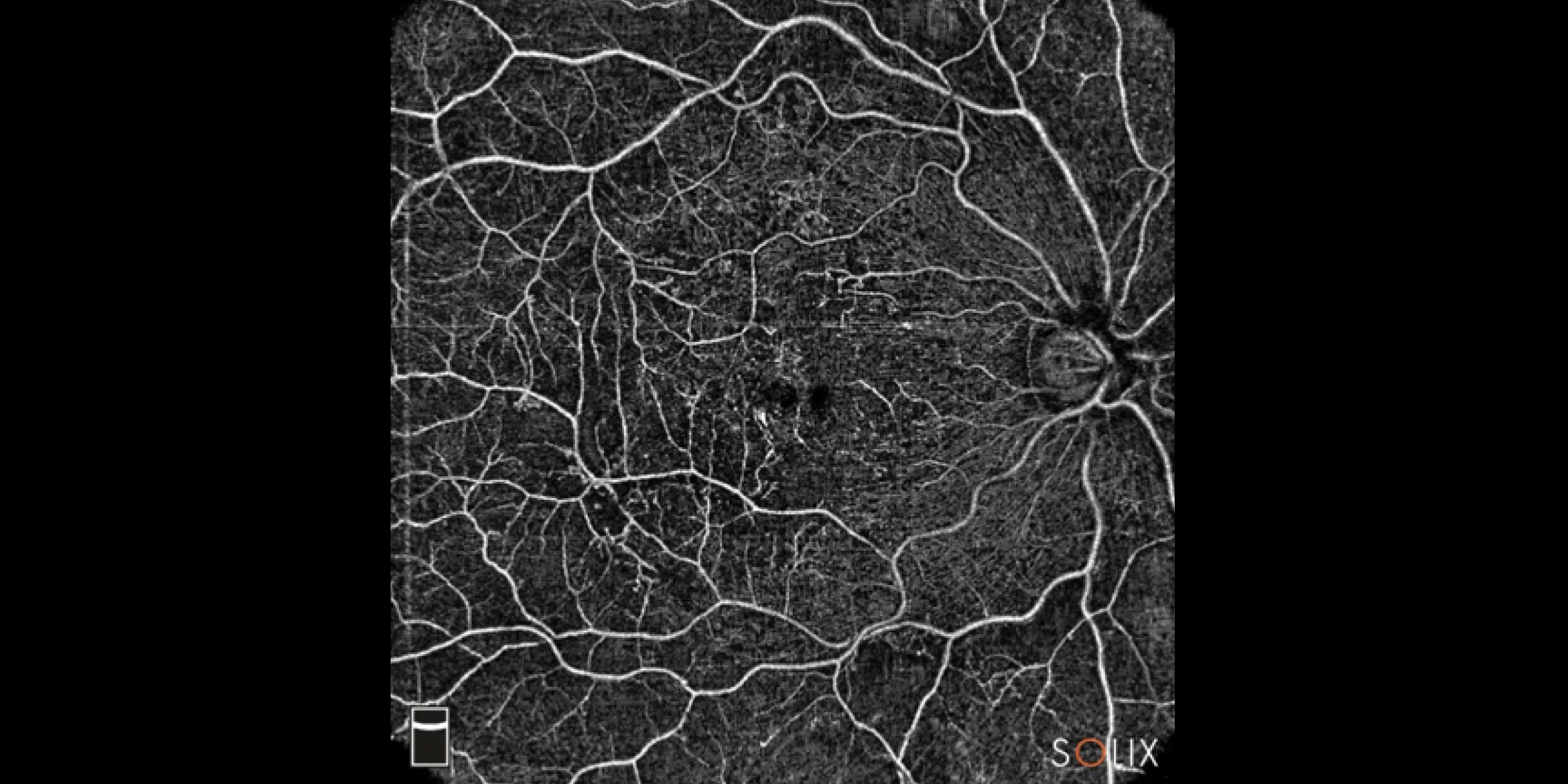
Ultra-fast spectral-domain (SD) technology produces a wide and deep field of view that does not compromise image resolution. Multi-volume merge technology averages four scan volumes to deliver high-density images with pristine clarity and 3D vessel rendering enables visualization of retinal vasculature and vascular connectivity.

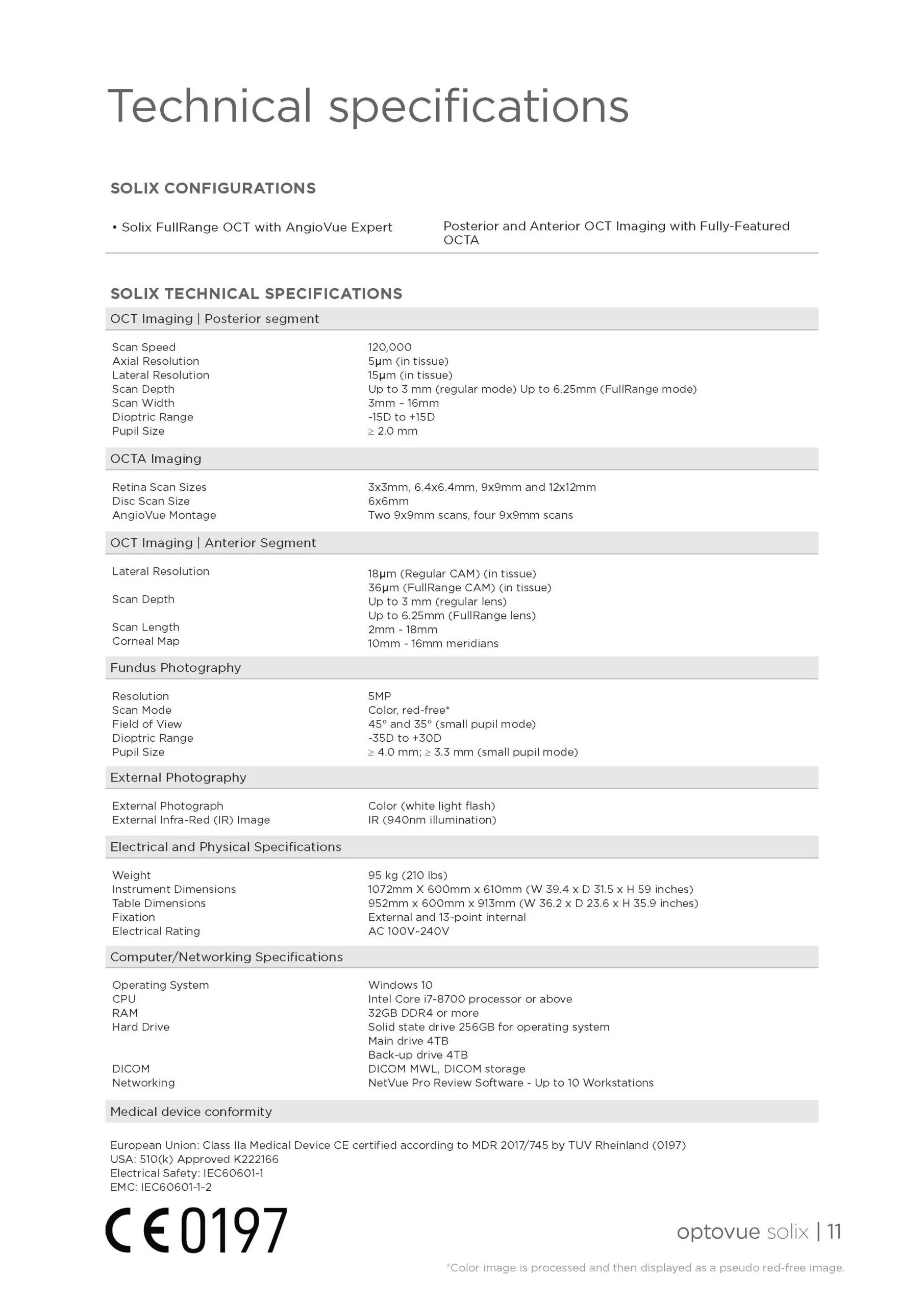
SOLIX FullRange anterior imaging provides stunning views of the entire anterior chamber from the front surface of the cornea to the anterior surface of the lens (18 x 6.5 mm) .

The integrated fundus camera provides both internal and external colour photography, combined with the IR imaging this comprehensive anterior segment package expands the clinical possibilities to address a broad range of patients.

Quantify epithelial, stromal and total corneal thickness with the 10mm Corneal Layer Map, which features 16 meridians to fully cover the LRS transition zone. The Highlight Tool to further appreciate subtle changes in epithelial thickness for early kerataconus detection. The Change Analysis report measures changes in thickness between visits.

OCT topography provides a comprehensive assessment of BOTH the anterior and posterior surfaces of the cornea while
being able to differentiate epithelial cells and stroma.


Screening for refractive surgery
Raise awareness on the importance of Epithelial Thickness Mapping (ETM) in refractive surgery screening.

Retina protocol
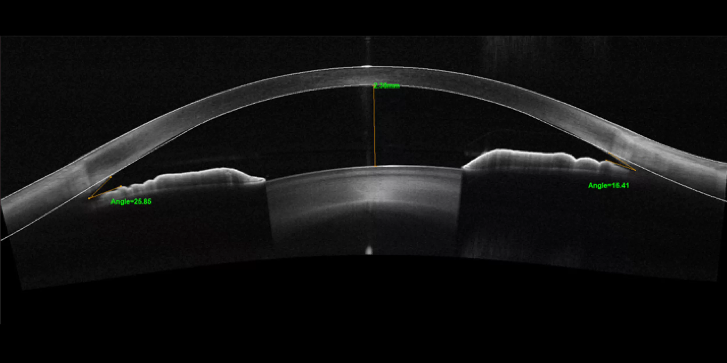
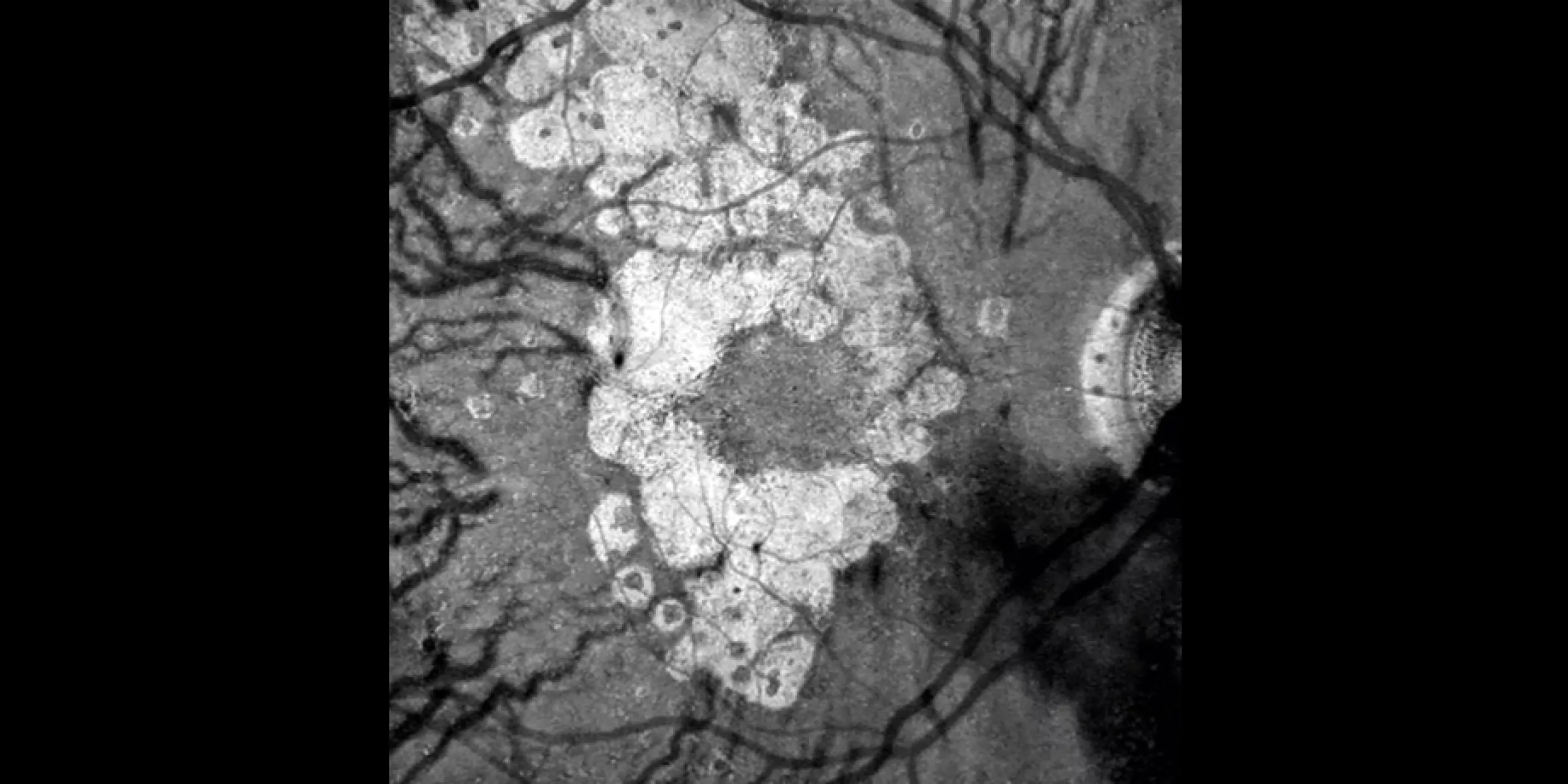
SOLIX delivers pristine images of retinal structures with unprecedented views of the vitreous and choroid, that enable confident diagnosis and management of retinal pathologies.
Protocolo Glaucoma
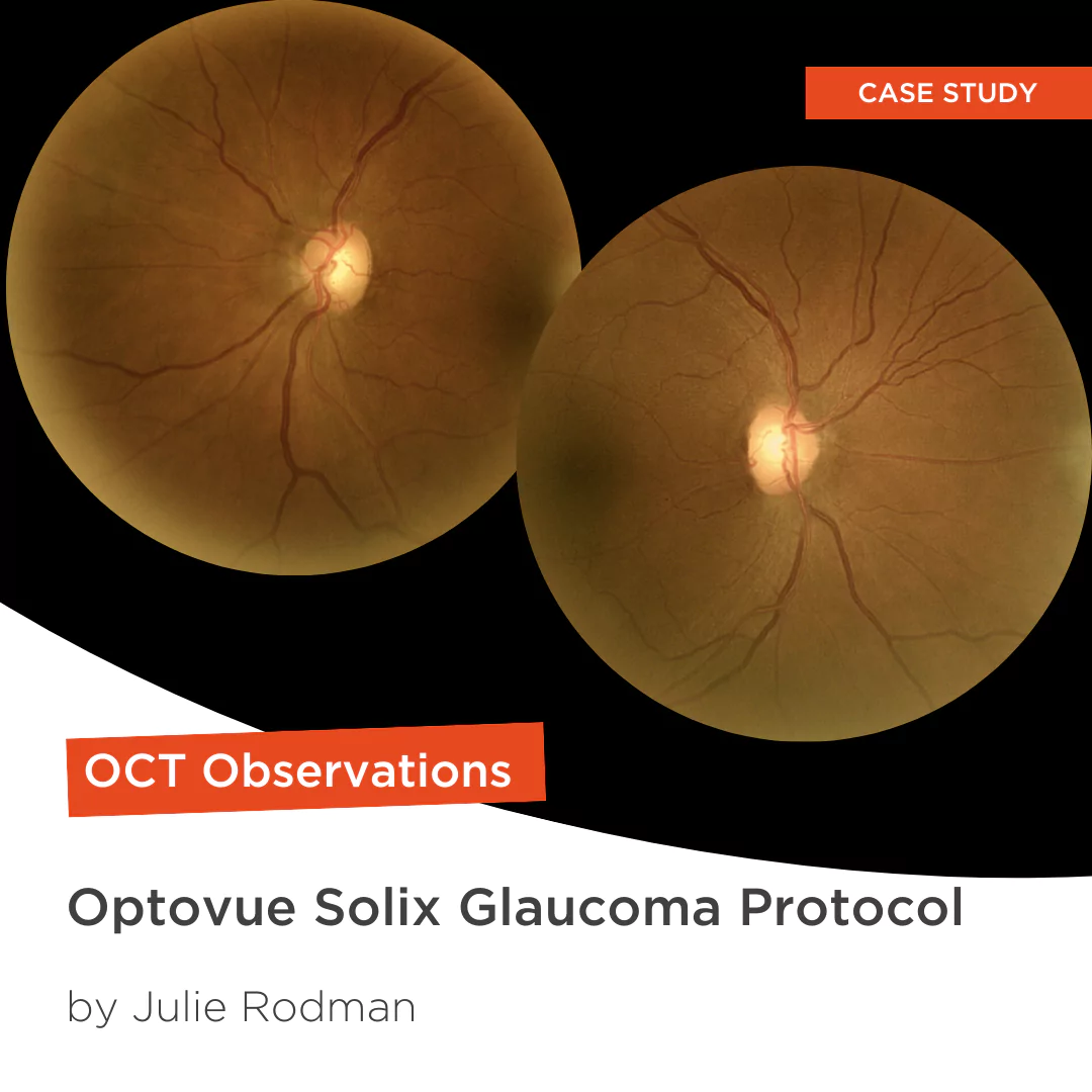
The SOLIX glaucoma package delivers in-depth analysis of the optic nerve head structure and vasculature. Optovue-exclusive scans bring additional insights that aid in clinical decision making.
Protocole Glaucome
The SOLIX glaucoma package delivers in-depth analysis of the optic nerve head structure and vasculature. Optovue-exclusive scans bring additional insights that aid in clinical decision making.
Glaucoma protocol
The SOLIX glaucoma package delivers in-depth analysis of the optic nerve head structure and vasculature. Optovue-exclusive scans bring additional insights that aid in clinical decision making.

We are big fans of Briot
Dr. Gray Sass, OD Wildwood Eyecare
Screening for refractive surgery
Raise awareness on the importance of Epithelial Thickness Mapping (ETM) in refractive surgery screening.
Retina protocol
SOLIX delivers pristine images of retinal structures with unprecedented views of the vitreous and choroid, that enable confident diagnosis and management of retinal pathologies.
Protocolo Glaucoma
The SOLIX glaucoma package delivers in-depth analysis of the optic nerve head structure and vasculature. Optovue-exclusive scans bring additional insights that aid in clinical decision making.
Protocole Glaucome
The SOLIX glaucoma package delivers in-depth analysis of the optic nerve head structure and vasculature. Optovue-exclusive scans bring additional insights that aid in clinical decision making.
Glaucoma protocol
The SOLIX glaucoma package delivers in-depth analysis of the optic nerve head structure and vasculature. Optovue-exclusive scans bring additional insights that aid in clinical decision making.
We are big fans of Briot
Dr. Gray Sass, OD Wildwood Eyecare

My office has had a Briot edger for over 10 years
Dr. Albert Morier Consumer Optical

”Implementing the Briot® Attitude 2 has been effortless. It saves me time so I don’t need to spend extra hours in the office.“
Dr. Douglas King Owner, Family Eye Care Optometry Center

Dr. Paul Karpecki Kentucky Eye Institute
Dr. Paul Karpecki Kentucky Eye Institute

Dr. John Gelles is the director of the specialty contact lens division at The Cornea and Laser Eye Institute – Hersh Vision Group and The CLEI Center for Keratoconus.
Dr. John Gelles is the director of the specialty contact lens division at The Cornea and Laser Eye Institute – Hersh Vision Group and The CLEI Center for Keratoconus.

“I can safely and efficiently practice while also giving the patient confidence they are receiving top notch and modern care.”
Jordan Jones, O.D. Director of Eyecare, Wear Eyewear