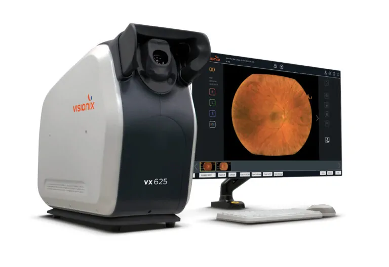
Capture crystal clear retinal images

Fundus camera

Fundus images, also known as retinographies, are pictures of the posterior part of the eye, including the retina, the optic nerve, and the blood vessels. They serve as a valuable tool for screening, diagnosing, and monitoring eye diseases.

Benefits of fundus images

Visionix

High resolution

Allows you to obtain a detailed view of the retina.

Icon

High sensitivity

Detection of retinal disorders at an early stage. Fundus images are sensitive to subtle changes in the retina.

Ease of interpretation

Authorizing healthcare professionals to provide rapid and accurate diagnoses.

Visionix expertise

Our fundus images offer high resolution, high sensitivity and a wide field of observation. They are obtained using state-of-the-art, fully automated equipment and proven technique.

Our fundus images are also essential for the detection of ocular pathologies. Vision professionals are experienced in interpreting fundus images and are able to provide accurate and comprehensive diagnoses.

Product

VX 610

  • Excellent image quality – The VX 610 camera utilises an innovative cross-polarized light technology that provides excellent image quality while maintaining a compact footprint.
     
  • Early automatic detect signs of more than 13 different retinal pathologies – The VX 610 Artificial Intelligence EyeWisdom®MCS solution covers 13 common retinal pathologies.With most retinal diseases, the primary goal of treatment is to slow disease progression and preserve the retina and associated vision.
     
  • No need to dilate the fundus through the pupil – Non-mydriatic retinograph that allows the observation, capture and recording of very high definition images without dilating the fundus through the pupil.
  • A.I. secures sensitivity and specificity – The ability of the A.I. to correctly tag a positive result is 93%. The ability of the A.I. to correctly tag a negative result is 90,6%
  • Automatic capture of high-quality images – For detailed screening and evaluation with minimal personal training
  • Modern gesture interaction – The intuitive interface facilitates a quick start and smooth navigation

Need more information?

Our teams are at your disposal to advise you

FAQ

What is VX 610?

The VX 610 is a non-mydriatic and automated fundus camera, easy to use and suitable for delegation. This camera allows capturing high-quality images under standard lighting conditions. Thanks to its compact and modern design, it fits perfectly into any examination room.

How does the Artificial Intelligence (A.I.) technology work with VX610?

The VX610 results can be transferred to a certified AI medical device to automate the analysis of fundus images. The technology is based on Deep Learning, a machine learning method which allows the computer to compare the images obtained to a bank of millions of images and thus detect anomalies.

What pathologies can be screened with A.I. technology based on VX610 images?

The A.I. medical solution using VX610 images addresses 13 retinal pathologies, including 4 major conditions which are dry age-related macular degeneration (non exudative AMD), wet AMD (exudative), suspected glaucoma and diabetic retinopathy.

What are the main advantages of a non-mydriatic camera?

A non-mydriatic camera allows the patient to avoid the unpleasant side effects of pupil dilation, such as blurred vision, sensitivity to light, and difficulty reading. Additionally, non-mydriatic fundus cameras are generally faster, less expensive, and easier to use than mydriatic fundus cameras.