


The VX 625 camera utilizes true color technology delivering retinal images with coloration that accurately represents the true appearance of the fundus.
Dedicated red, green, and blue channels for layer-specific assessment of the retina

The VX 625 is a ultra-widefield retinal camera designed to capture and record high-resolution images spanning 176 degrees of the retina in a single acquisition without the need for dilating the pupil

The VX 625 features a highly intuitive interface, allowing clinicians and staff to operate the system confidently with minimal training requirements

User friendly software for efficient documentation and patient monitoring
Clinicians can optimise visualisation with:

The VX 625 delivers true-color, ultra-widefield retinal images up to 176° in a single acquisition, allowing clinicians to view far beyond the traditional 30–45° field. With automated montage, the field of view can be expanded to approximately 220° using two images and to more than 260° with four images, allowing assessment of over 95% of the retina

Field of view : 176° (5% tolerance)
Field of view with mosaic function : 220° with 2 images
Retinal resolution : 14 microns?
Fixations : 5 standard up to many
Weight : 21 kg
Size : 250 x 500 x 470mm


Screening for refractive surgery
Raise awareness on the importance of Epithelial Thickness Mapping (ETM) in refractive surgery screening.

Retina protocol
SOLIX delivers pristine images of retinal structures with unprecedented views of the vitreous and choroid, that enable confident diagnosis and management of retinal pathologies.
Protocolo Glaucoma
The SOLIX glaucoma package delivers in-depth analysis of the optic nerve head structure and vasculature. Optovue-exclusive scans bring additional insights that aid in clinical decision making.
Protocole Glaucome
The SOLIX glaucoma package delivers in-depth analysis of the optic nerve head structure and vasculature. Optovue-exclusive scans bring additional insights that aid in clinical decision making.
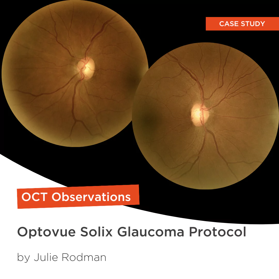
Glaucoma protocol
The SOLIX glaucoma package delivers in-depth analysis of the optic nerve head structure and vasculature. Optovue-exclusive scans bring additional insights that aid in clinical decision making.

We are big fans of Briot
Dr. Gray Sass, OD Wildwood Eyecare
Screening for refractive surgery
Raise awareness on the importance of Epithelial Thickness Mapping (ETM) in refractive surgery screening.
Retina protocol
SOLIX delivers pristine images of retinal structures with unprecedented views of the vitreous and choroid, that enable confident diagnosis and management of retinal pathologies.
Protocolo Glaucoma
The SOLIX glaucoma package delivers in-depth analysis of the optic nerve head structure and vasculature. Optovue-exclusive scans bring additional insights that aid in clinical decision making.
Protocole Glaucome
The SOLIX glaucoma package delivers in-depth analysis of the optic nerve head structure and vasculature. Optovue-exclusive scans bring additional insights that aid in clinical decision making.
Glaucoma protocol
The SOLIX glaucoma package delivers in-depth analysis of the optic nerve head structure and vasculature. Optovue-exclusive scans bring additional insights that aid in clinical decision making.
We are big fans of Briot
Dr. Gray Sass, OD Wildwood Eyecare

My office has had a Briot edger for over 10 years
Dr. Albert Morier Consumer Optical

”Implementing the Briot® Attitude 2 has been effortless. It saves me time so I don’t need to spend extra hours in the office.“
Dr. Douglas King Owner, Family Eye Care Optometry Center

Dr. Paul Karpecki Kentucky Eye Institute
Dr. Paul Karpecki Kentucky Eye Institute

Dr. John Gelles is the director of the specialty contact lens division at The Cornea and Laser Eye Institute – Hersh Vision Group and The CLEI Center for Keratoconus.
Dr. John Gelles is the director of the specialty contact lens division at The Cornea and Laser Eye Institute – Hersh Vision Group and The CLEI Center for Keratoconus.

“I can safely and efficiently practice while also giving the patient confidence they are receiving top notch and modern care.”
Jordan Jones, O.D. Director of Eyecare, Wear Eyewear