


With just a single tap, the Optovue Autofusion can automatically align, focus, track, capture images, and provide measurement results for both Macular OCT and Disc OCT.
Additionally, when the optional CAM accessory is attached, it performs auto measurement for Pachymetry and angles. Utilizing 3D tracking, focusing technology and auto capture of fundus images, the Optovue Autofusion streamlines the
examination process for doctors while enhancing patient care.

Optovue Autofusion offers optic nerve and macular analysis from a single 3D Wide scan. Individual optic nerve scan, provides progressive RNFL, and ONH parameter evaluation. 3D Macula includes progressive ETDRS THICKNESS maps, and GCC analysis. Other posterior reports include 5-Line Cross, Line , Wide Line mode and Radial scans. Anterior reports include Corneal thickness, Angles. Detailed and pre-formatted reports can be easily exported, printed or shared.

Este informe de seguimiento resalta los cambios estructurales entre los exámenes, proporcionando una evaluación detallada y cuantitativa tanto del disco óptico como de la capa de fibras nerviosas de la retina (RNFL). Los parámetros clave incluyen: Cambio en el grosor de la RNFL (OD-OS); Análisis de tendencia de la RNFL (OD-OS); Mapa de significancia de la RNFL (μm); Topografía del disco. Este informe completo combina datos cuantitativos y visuales para mejorar la detección de cambios tempranos, convirtiéndolo en una herramienta vital en el manejo clínico de las patologías del nervio óptico, especialmente en el seguimiento del glaucoma.

The Optovue Autofusion offers outstanding performance in anterior segment imaging, combining high-resolution scans with dedicated analysis tools for precise evaluation of corneal structures, and angle measurements for surgical planning. Its advanced anterior segment module enhances diagnostic confidence and broadens clinical applications, making it an indispensable tool for comprehensive anterior segment assessment.

NOT FOR THE USA
OCT SCANNING FUNCTION
FUNDUS CAMERA FUNCTION
GENERAL FUNCTION


Screening for refractive surgery
Raise awareness on the importance of Epithelial Thickness Mapping (ETM) in refractive surgery screening.

Retina protocol
SOLIX delivers pristine images of retinal structures with unprecedented views of the vitreous and choroid, that enable confident diagnosis and management of retinal pathologies.
Protocolo Glaucoma
The SOLIX glaucoma package delivers in-depth analysis of the optic nerve head structure and vasculature. Optovue-exclusive scans bring additional insights that aid in clinical decision making.
Protocole Glaucome
The SOLIX glaucoma package delivers in-depth analysis of the optic nerve head structure and vasculature. Optovue-exclusive scans bring additional insights that aid in clinical decision making.
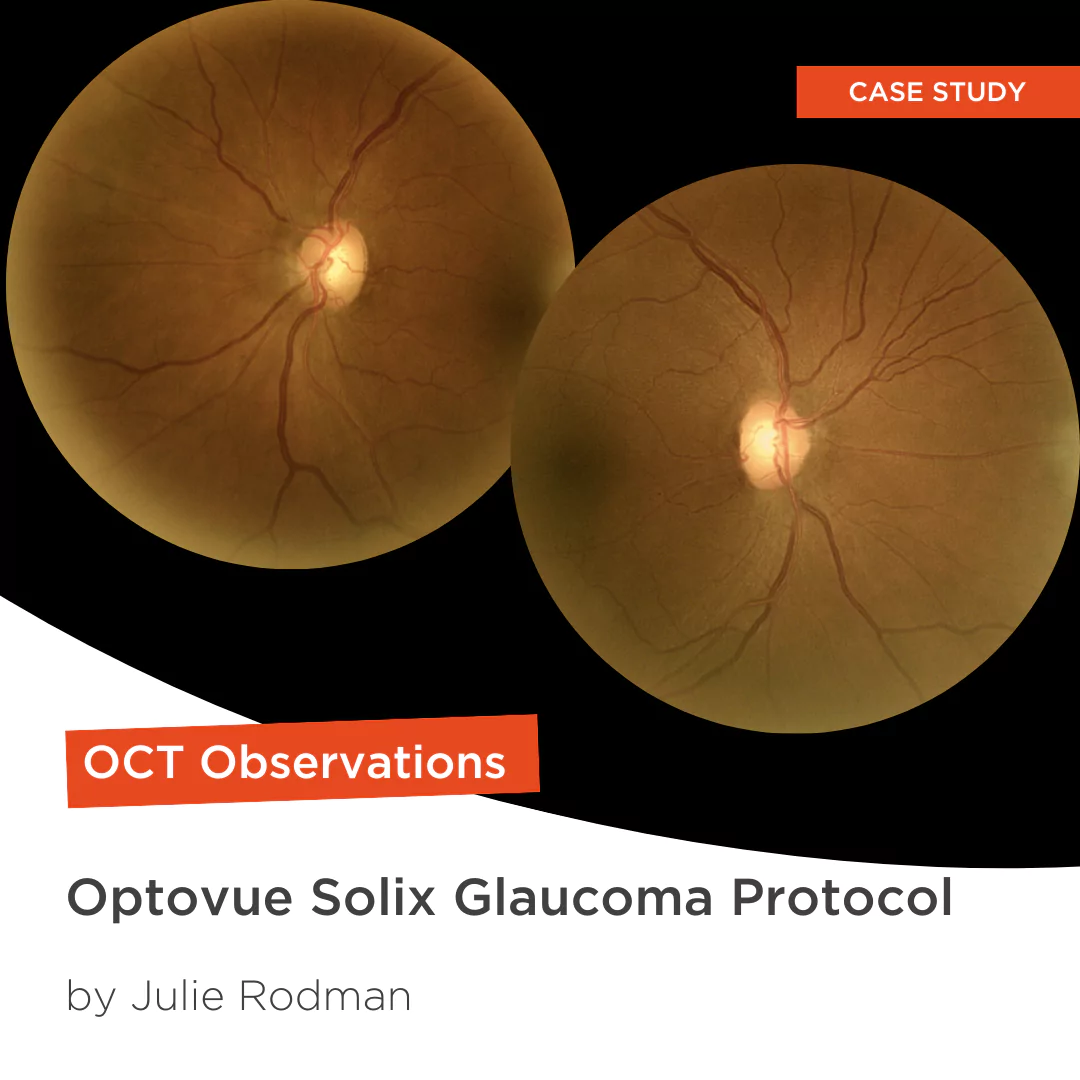
Glaucoma protocol
The SOLIX glaucoma package delivers in-depth analysis of the optic nerve head structure and vasculature. Optovue-exclusive scans bring additional insights that aid in clinical decision making.

We are big fans of Briot
Dr. Gray Sass, OD Wildwood Eyecare
Screening for refractive surgery
Raise awareness on the importance of Epithelial Thickness Mapping (ETM) in refractive surgery screening.
Retina protocol
SOLIX delivers pristine images of retinal structures with unprecedented views of the vitreous and choroid, that enable confident diagnosis and management of retinal pathologies.
Protocolo Glaucoma
The SOLIX glaucoma package delivers in-depth analysis of the optic nerve head structure and vasculature. Optovue-exclusive scans bring additional insights that aid in clinical decision making.
Protocole Glaucome
The SOLIX glaucoma package delivers in-depth analysis of the optic nerve head structure and vasculature. Optovue-exclusive scans bring additional insights that aid in clinical decision making.
Glaucoma protocol
The SOLIX glaucoma package delivers in-depth analysis of the optic nerve head structure and vasculature. Optovue-exclusive scans bring additional insights that aid in clinical decision making.
We are big fans of Briot
Dr. Gray Sass, OD Wildwood Eyecare

My office has had a Briot edger for over 10 years
Dr. Albert Morier Consumer Optical

”Implementing the Briot® Attitude 2 has been effortless. It saves me time so I don’t need to spend extra hours in the office.“
Dr. Douglas King Owner, Family Eye Care Optometry Center

Dr. Paul Karpecki Kentucky Eye Institute
Dr. Paul Karpecki Kentucky Eye Institute

Dr. John Gelles is the director of the specialty contact lens division at The Cornea and Laser Eye Institute – Hersh Vision Group and The CLEI Center for Keratoconus.
Dr. John Gelles is the director of the specialty contact lens division at The Cornea and Laser Eye Institute – Hersh Vision Group and The CLEI Center for Keratoconus.

“I can safely and efficiently practice while also giving the patient confidence they are receiving top notch and modern care.”
Jordan Jones, O.D. Director of Eyecare, Wear Eyewear