


iScan 80 OCT offers the scans available on a traditional OCT with the added benefit of simplified operation. Scan acquisition is as easy as positioning the patient with the assistance of iScan 80’s Pupil Alignment Technology, choosing the scan and pushing start.

Scan acquisition is as easy as positioning the patient with the assistance of iScan 80’s Pupil Alignment Technology, choosing the scan and pushing start. iScan 80 performs all of the focus and alignment operations while talking the patient through the entire exam.

Compact and portable, iscan 80’s streamlined design integrates the operator interface, display, patient interface and scan head into one console that can be moved from station to station and mounted on any tabletop. Then comes the easy part—plug in, switch on and start scanning. It does not require a dark exam area.

Take a new approach to protecting your patients’ eye health with the iWellness Exam, available from Optovue. This quick and easy scan gives you a detailed look at the health of your patient’s retina and ganglion cell complex with robust normative comparison, to help you identify signs of disease before they may be noticeable on clinical exam. And because the iWellness scan is separate from the iScan 80 medically-billable scans, it provides a new revenue stream for the practice.
iScan 80 is available in two configurations to give you flexibility in incorporating OCT. iScan 80 Essential offers the iWellness scan as well as basic retina and glaucoma scans for practices who are new to OCT technology. For practices seeking a fully-featured system, iScan 80 Comprehensive provides iWellness along with all of the retina, optic disc and anterior segment scans available on the iVue system.

| Feature | Value |
|---|---|
| OCT Image 80,000 A-scan/second | • |
| Depth Resolution (in tissue) 5.0 μm | • |
| Transverse Resolution 15 μm (retina) | • |
| . | Value |
|---|---|
| Depth 2 – 2.3mm (retina) | • |
| Scan Beam Wavelength 840nm (+/-10nm) | • |
| . | Value |
|---|---|
| FOV | (H) 21° x (V) 21° |
| Minimum Pupil Diameter | 2mm |
| External Image (Live IR) FOV | 13mm x 8mm |
| . | Value |
|---|---|
| Dimensions (in.) | (W) 19.81 x (L) 15.83 x (H) 17.82 |
| Weight (lb.) | 42.94 |
| . | Value |
|---|---|
| Operating System | Win 10 – 64 bit |
| Processor Speed | Intel® Celeron (i7-8700T 2.40 GHz) |
| Network Bandwidth | 1Gbps or higher |
| Computer RAM | 16 GB or higher |
| Monitor Resolution | 24 in.1920×1080 60Hz |
| DICOM compatible | • |


Screening for refractive surgery
Raise awareness on the importance of Epithelial Thickness Mapping (ETM) in refractive surgery screening.

Retina protocol
SOLIX delivers pristine images of retinal structures with unprecedented views of the vitreous and choroid, that enable confident diagnosis and management of retinal pathologies.
Protocolo Glaucoma
The SOLIX glaucoma package delivers in-depth analysis of the optic nerve head structure and vasculature. Optovue-exclusive scans bring additional insights that aid in clinical decision making.
Protocole Glaucome
The SOLIX glaucoma package delivers in-depth analysis of the optic nerve head structure and vasculature. Optovue-exclusive scans bring additional insights that aid in clinical decision making.
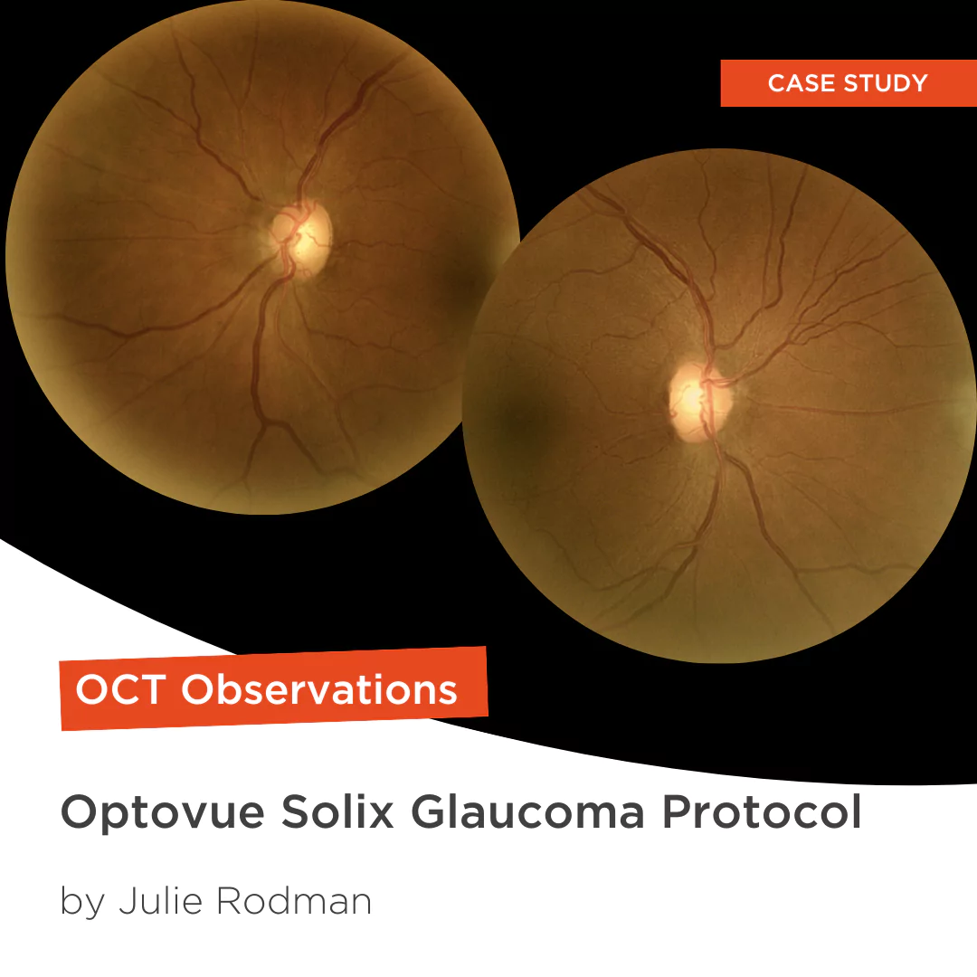
Glaucoma protocol
The SOLIX glaucoma package delivers in-depth analysis of the optic nerve head structure and vasculature. Optovue-exclusive scans bring additional insights that aid in clinical decision making.

We are big fans of Briot
Dr. Gray Sass, OD Wildwood Eyecare
Screening for refractive surgery
Raise awareness on the importance of Epithelial Thickness Mapping (ETM) in refractive surgery screening.
Retina protocol
SOLIX delivers pristine images of retinal structures with unprecedented views of the vitreous and choroid, that enable confident diagnosis and management of retinal pathologies.
Protocolo Glaucoma
The SOLIX glaucoma package delivers in-depth analysis of the optic nerve head structure and vasculature. Optovue-exclusive scans bring additional insights that aid in clinical decision making.
Protocole Glaucome
The SOLIX glaucoma package delivers in-depth analysis of the optic nerve head structure and vasculature. Optovue-exclusive scans bring additional insights that aid in clinical decision making.
Glaucoma protocol
The SOLIX glaucoma package delivers in-depth analysis of the optic nerve head structure and vasculature. Optovue-exclusive scans bring additional insights that aid in clinical decision making.
We are big fans of Briot
Dr. Gray Sass, OD Wildwood Eyecare

My office has had a Briot edger for over 10 years
Dr. Albert Morier Consumer Optical

”Implementing the Briot® Attitude 2 has been effortless. It saves me time so I don’t need to spend extra hours in the office.“
Dr. Douglas King Owner, Family Eye Care Optometry Center

Dr. Paul Karpecki Kentucky Eye Institute
Dr. Paul Karpecki Kentucky Eye Institute

Dr. John Gelles is the director of the specialty contact lens division at The Cornea and Laser Eye Institute – Hersh Vision Group and The CLEI Center for Keratoconus.
Dr. John Gelles is the director of the specialty contact lens division at The Cornea and Laser Eye Institute – Hersh Vision Group and The CLEI Center for Keratoconus.

“I can safely and efficiently practice while also giving the patient confidence they are receiving top notch and modern care.”
Jordan Jones, O.D. Director of Eyecare, Wear Eyewear