FDA clears first ever FullRange® Spectral Domain -OCT/OCT-A – Optovue Solix by Visionix
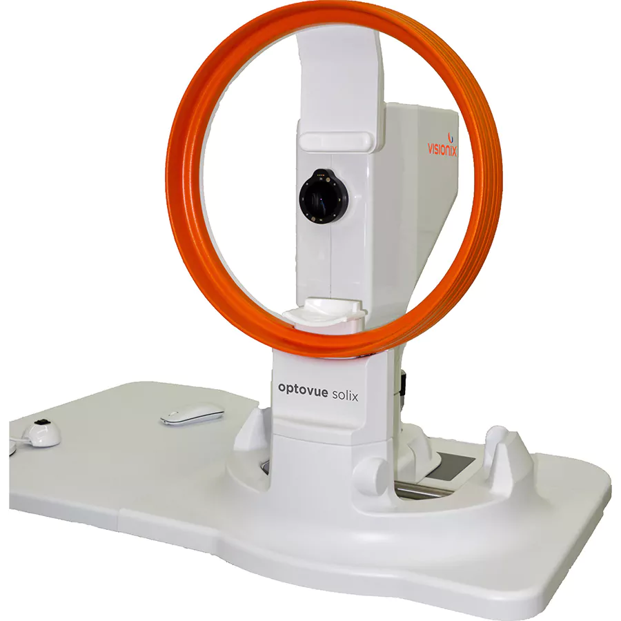
SOLIX is new technology built upon a proven foundation of high-speed Spectral Domain OCT. This FullRange® platform empowers practitioners to identify and manage numerous pathologies from the front of the eye to the back for a vastly superior diagnostic experience. SOLIX delivers multiple tools for a new generation of disease management that improves throughput and enables superior patient care.
FullRange single scan imaging shows entire anterior chamber from the front of the cornea to the anterior surface of the lens or entire Crystalline lens.
New advanced scans and glaucoma analytics take glaucoma scanning to the next level incorporating Dual Track, SSADA, MCT, and AI segmentation with repeatability and reproducibility 2 times better than before.
Wellness capabilities that have become part of a new standard of care for patients suspected of both retinal pathologies and/or glaucoma. The AngioWellness scan enables comprehensive assessment of your diabetic patients and glaucoma suspects by combining structural information on retinal and ganglion cell thickness with objective metrics on retinal vasculature. Utilize FAZ Analytics to uncover early indicators of diabetic changes.
Optovue Solix is a new technology built upon a proven foundation of high-speed Spectral Domain OCT. With 120kHz scan speed, higher scan density, and better precision this FullRange OCT delivers unparalleled data and efficiency.

The Solix platform is available in two configurations that are easily upgradeable, so your OCT system meets the needs of your practice today and into the future.
SOLIX CONFIGURATIONS
SOLIX TECHNICAL SPECIFICATIONS
OCT Imaging | Retina
OCT-A Imaging
OCT Imaging | Anterior Segment
Fundus Photography
External Photography
Electrical and Physical Specifications
Computer/Networking Specifications
Solix |
Solix Essential |
iFusion 80 |
iScan 80 |
iVue 80 |
|
Technology |
|||||
| Transverse Resolution(15μm) | ✔️ | ✔️ | ✔️ | ✔️ | ✔️ |
| Scan Speed | 120kHz | 120kHz | 80kHz | 80kHz | 80kHz |
| Axial Resolution(5μm) | ✔️ | ✔️ | ✔️ | ✔️ | ✔️ |
| iWellness scan | ✔️ | ✔️ | ✔️ | ✔️ | ✔️ |
| AngioVue single scan for structural & vascular OCT | ✔️ | ✔️ | |||
| AngioVue OCT-A with enhanced metrics | ✔️ | ✔️ | |||
| Multi-volume averaging, SSADA | ✔️ | ✔️ | |||
| 3D PAR 2.0 | ✔️ | ✔️ | |||
| DualTrac – Motion Correction Technology (MCT) | ✔️ | ✔️ | |||
| Pixel x Pixel deviation mapping | ✔️ | ✔️ | |||
| AngioWellness scan | ✔️ | ✔️ | |||
| Fully Automated with voice guided technology | ✔️ | ||||
Anterior Segment |
|||||
| Anterior Radial (12mm) | ✔️ | ✔️ | ✔️ | ✔️ | ✔️ |
| Angle scan | ✔️ | ✔️ | ✔️ | ✔️ | ✔️ |
| Epithelial, stromal, and corneal, thickness mapping | ✔️ | ✔️ | ✔️ | ✔️ | ✔️ |
| Pachymetry | 10mm | 10mm | 6mm | 6mm | 6mm |
| Angle scan and analysis with 4-up display | ✔️ | ✔️ | ✔️ | ✔️ | ✔️ |
| External color camera | ✔️ | ✔️ | |||
| FullRange Anterior segment 18 x 6.25mm | ✔️ | ||||
| Exterior IR lid imaging | ✔️ | ||||
Glaucoma |
|||||
| 3D Disc Cube | ✔️ | ✔️ | ✔️ | ✔️ | ✔️ |
| GCC Analysis | ✔️ | ✔️ | ✔️ | ✔️ | ✔️ |
| Nerve Fiber Layer Analysis | ✔️ | ✔️ | ✔️ | ✔️ | ✔️ |
| Comprehensive single eye and OU Reports | ✔️ | ✔️ | ✔️ | ✔️ | ✔️ |
| RPC density map and values | ✔️ | ✔️ | |||
| 100 μm RNFL circle at 3.45mm | ✔️ | ✔️ | |||
| 3 Times Repeatablity and Reproducability (R&R) | ✔️ | ✔️ | |||
Retina |
|||||
| EnFace | 12 x 12 mm | 12 x 12mm | 12 x 9mm | 12 x 9mm | 12 x 9mm |
| 3D Retina Cube | ✔️ | ✔️ | ✔️ | ✔️ | ✔️ |
| Radial Line | ✔️ | ✔️ | ✔️ | ✔️ | ✔️ |
| 512 OCT-A scans | ✔️ | ✔️ | |||
| 3D vessel rendering | ✔️ | ✔️ | |||
| AngioVue QuadMontage | ✔️ | ✔️ | |||
| Retinal Thickness Map (Widefield OCT-A 12 x 12mm, 9 x 9mm) | ✔️ | ✔️ | |||
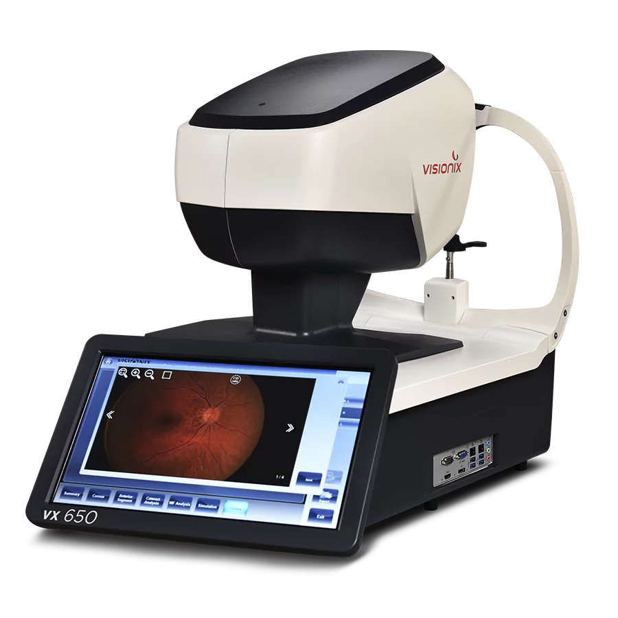
| Fundus Camera | ✔️ | ✔️ | |||
| FullRange Retinal scan 16 x 6.25mm | ✔️ | ||||

Screening for refractive surgery
Raise awareness on the importance of Epithelial Thickness Mapping (ETM) in refractive surgery screening.

Retina protocol
SOLIX delivers pristine images of retinal structures with unprecedented views of the vitreous and choroid, that enable confident diagnosis and management of retinal pathologies.

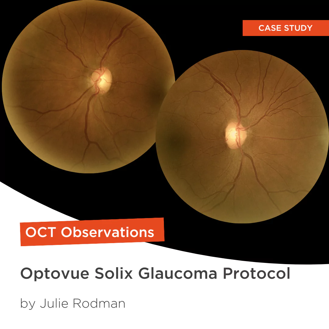
Glaucoma protocol
The SOLIX glaucoma package delivers in-depth analysis of the optic nerve head structure and vasculature. Optovue-exclusive scans bring additional insights that aid in clinical decision making.
Description / Lorem ipsum dolor sit amet consectetur. Arcu dolor suspendisse commodo a faucibus amet. Consequat lacinia diam eget imperdiet placerat. Massa sed neque pharetra quis interdum egestas mi.


