


Revenio’s subsidiary, iCare and Visionix are joining forces to create a stronger and more comprehensive eye care solutions provider, bringing together two highly complementary companies with shared ambitions for innovation, connectivity, and customer value.
Together, the two companies are expanding capabilities across diagnostics, imaging, refraction, OCT, and workflow solutions while continuing to support customers and partners as usual.
The combination strengthens both companies’ ability to support optical retail, optometry, and ophthalmology with connected technologies, integrated workflows, and advanced diagnostic solutions across the full patient journey.
Customers and partners will benefit from:
Importantly, existing customer contacts, support channels, and operations remain unchanged.
Together, iCare and Visionix are building the future of connected eye care.
The VX 40 Pro is the next-generation solution that streamlines the verification of frames and lenses, providing opticians with an easy-to-use, turnkey device that ensures accurate, fast, and reliable results.


Add an extra layer of security to your verification process, ensuring more accurate results and reducing returns and customer dissatisfaction.

The device checks spherical power, cylinder, axis, prisms, and optical center alignment.

See all measurements directly on the screen at true 1:1 scale, making verification intuitive and precise.

Automatically generate a comprehensive report for every lens inspection, ensuring full traceability and compliance for your records and your customers


Comprehensive Verification in 30 Seconds
Verify lens geometry, power, mapping, pupillary distance, height, and prismatic effect with just one click.
No more manual measurements. Save time and eliminate errors.
The VX 40 Couture delivers unmatched accuracy, ensuring your optical frames are perfectly matched to the customer’s medical prescription.
Integrated with Briot Couture edging system for seamless compatibility and enhanced precision.
After verification, the system generates a compliance report, providing tangible proof of conformity for every assembly.
When connected to the full system, the VX 40 Couture offers complete automation, making the verification process faster, more accurate, and more reliable.
Compatible with Visionix edging systems
Connected to the Briot Couture or Weco E7 edging system, the VX 40 Couture allows the optician to verify several essential criteria with a single click.


” Since integrating the VX 40 Pro into our workflow, we’ve experienced a noticeable improvement in efficiency and quality control. The system is incredibly easy to use, and the ability to generate compliance reports has been a game changer.”
Request a demo
Request a quote
Contact us today to learn more about how the VX 40 Pro can transform your optical workflow and guarantee quality with every frame.
![]()

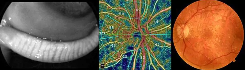
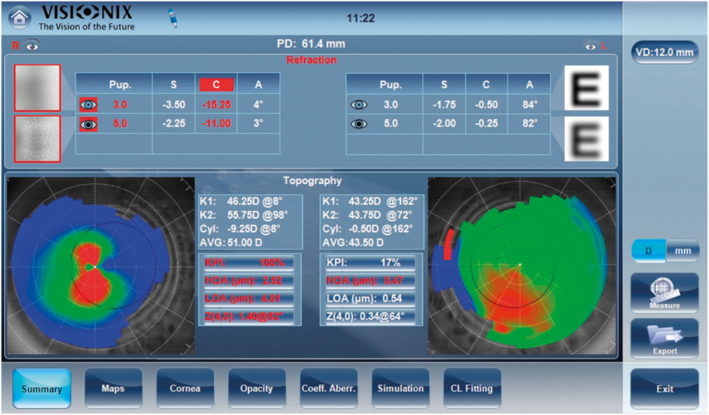
The Visionix VX 118 gives practitioners the comprehensive clinical insights needed to screen, evaluate, and monitor a wide range of various eye conditions, including keratoconus and cataracts. With its comprehensive suite of tools and advanced technologies, it provides practitioners with the necessary insights to deliver optimal patient care.
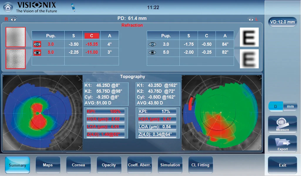
For keratoconus screening and monitoring, the VX 118 employs topography maps, axial, tangential elevation, and refraction maps, allowing for detailed analysis of corneal shape and irregularities. The device also features the Keratoconus Probability Index (KPI) and enables ongoing monitoring of keratoconus progression. Additionally, internal astigmatism measurement, eccentricity, meridian tables, and corneal aberrometry further enhance diagnostic accuracy.
Utilizing wavefront analysis with Shack-Hartmann technology, Placido rings, and Scheimpflug imaging, the VX 118 ensures precise evaluation of corneal abnormalities associated with keratoconus, enabling early detection and intervention.
For cataract assessment, the VX 118 enables visualization of crystalline opacities and analysis of wavefront aberrations, allowing practitioners to differentiate between corneal and lenticular/internal aberrations. Additional capabilities include internal astigmatism measurement, kappa angle evaluation for intraocular lens (IOL) centering, Z.4.0 value assessment for aspheric implant selection, and lens opacity classification using LOCS II and III scales. Combining Scheimpflug imaging, retroillumination, Shack-Hartmann technology, and Placido rings, the VX 118 delivers comprehensive insight into cataract severity and supports confident treatment planning.
The Visionix VX 118 is designed to make a meaningful impact in diagnostic screening. With fully automated, end-to-end capabilities, it handles a wide range of functions essential to comprehensive eye examinations. From complete refraction to the identification and monitoring of conditions such as keratoconus and cataracts, plus specialty contact lens fitting. It’s a versatile, all-in-one solution built for the demands of high-performing optometry and ophthalmology practices.
Complete refraction with objective measurements
A core strength of the VX 118 is its ability to deliver objective refraction measurements tailored to both day and night vision needs. The device performs objective refraction under both mesopic and photopic conditions, allowing practitioners to differentiate between a patient’s vision requirements across varying lighting environments. With 1,300 points analyzed across a 7mm diameter pupil, the VX 118 delivers highly detailed, accurate assessments of visual acuity and quality of vision, even on pupils as small as 1.2mm.
Differentiation between day and night vision needs
Objective refraction under mesopic and photopic conditions
Cataract assessment and monitoring
Keratoconus screening and monitoring
Fully automatic 3D and right/left eye alignments
Measurement of lower-order and higher-order aberrations
Comprehensive topography maps for corneal analysis
Scheimpflug imaging and Placido rings for precise evaluation

Request a demo
Request a quote
https://www.visionix.com/wp-content/uploads/2024/12/EN-Animation-page-show-case-1-2.mp4
Since its launch in 2012, this machine has transformed industry standards, combining simplicity, performance, and cutting-edge technology. Discover the evolution of this iconic machine through its three major milestones, each marked by significant advancements for users.
Launched in 2012, the Briot Emotion revolutionized the edging sector with its all-in-one concept. Designed to make the optical lens edging step more accessible and less intimidating, this edger combined ergonomics with ease of use. Thanks to its intuitive design and simplified functions, it enabled professionals and beginner opticians to carry out their edging tasks with great ease, without compromising on quality.

Launched in 2018, the Briot Emotion 2 made a giant step forward. This improved version integrated Gravitech technology, a patented innovation that offers high-precision optical tracing with a 1:1 ratio, ensuring the machine precisely follows the contours of the lens without distortion.

With the Briot Emotion 3, the new advanced anti-slippage edging cycle keeps the ophthalmic lens perfectly stable during edging, eliminating any risk of misalignment or damage. Combined with the double-sided pads recommended by Visionix, it offers increased stability and precision. The adjustable edging cycle speed allows secure work on high-value lenses, ensuring flawless finishes and optimal control.
Dive back into the essence of optical craftsmanship.
Imagine a time when every movement mattered, when expert hands carefully shaped each frame. With Visionix, this heritage lives on through our high-precision edgers, essential devices that redefine the excellence of optical assembly.
✨ With our Briot range of lens finishing equipment, our lens edgers do more than just edge, finish, and drill: they become the soul of the workshop, ensuring a perfect finish and incredible turn-around time, placing the customer first.
Let’s transform each of our assemblies into a technical masterpiece. Let’s consider our grinder as the nerve center of our store. 👓

Request a quote
Request a demo


Simple
Easy handling and formidable efficiency, allowing anyone to achieve quality results with minimal effort. The Briot Emotion quickly became a popular choice, solidifying its place among the most sought-after edgers.
Fast
Faster and more precise form capturing, increasing productivity while maintaining optimal result quality.
Precise
Ultra-precise and fast edging, enabling professionals to work confidently with high-end lenses while optimizing performance and safety. The Briot Emotion 3 has become the ultimate choice for demanding professionals who want to control their workshop.
Briot Emotion combines cutting-edge technology with a deep commitment to precision and quality. With over 12 years of experience, it ensures the highest standards in optical lens processing. Our tools are designed for professionals who demand excellence, offering reliable performance, innovation, and unmatched accuracy.
With over 12 years of experience, the Briot Emotion series is established as a leader in ophthalmic lens processing equipment, offering a track record of reliability and innovation.
Our engineers and experts continuously refine our technology, ensuring that each product meets the highest industry standards.
Briot Emotion devices are built with high-quality materials, ensuring durability and consistent performance over time.
Our products undergo rigorous testing to ensure they can withstand the wear and tear of daily use without compromising on quality.
Enhancing Every Lens Fitting with Precision and Care
Our edgers offer exceptional precision, ensuring that every lens is crafted to perfection for an impeccable fit in its frame.
The technology used in Briot Emotion devices enables faster, more accurate processing that reduce errors and enhance overall efficiency.
Our products are designed to put the technician’s skill at the forefront, providing lens finishing that supports expertise and elevates the craftsmanship of every optical fitting.


Featuring:
Pick up to six different maps to view at once. View any combination of anterior/posterior axial, tangential, and elevation topography maps with epithelial thickness, pachymetry, and much more!
Utilize Zernike data to optimize screening for laser procedures, IOLs, ICLs, and specialized contact lens selection. Enhance patient satisfaction by understanding post-treatment outcomes.


View side-by-side pachymetry, epithelial thickness, and stroma maps. Easily differentiate stromal and epithelial cells.
Pachymetry, epithelial thickness, and stroma trend analysis are available for assessing change and demonstrating your patient’s progress before and after treatment or surgery. Be able to detect and track keratoconic changes.


The FullRange® anterior chamber scan now includes auto-caliper placement.
“As our treatments for ocular conditions are becoming more readily available, it is critical that eyecare providers have the appropriate technology to maintain quality patient care. Often this means finding additional space for more and more devices. The addition of topography software module upgrade on the Optovue Solix device offers an accurate, easy to use solution to this problem. Our clinic can now obtain accurate topographic information faster than with any other topographer at our disposal, and we are able to do so at the same machine we image for screening of retinal conditions and optic nerve anomalies. The amount of data given by the Optovue Solix Topography module is unmatched. In a single second we can obtain information regarding the anterior corneal surface, posterior corneal surface, corneal thickness, and epithelial mapping all of which help us better serve our patients and ensure the highest level of patient care while maintaining clinical time and space efficiency.”
 Nathan Lighthizer, OD, FAAO
Associate Professor and Associate Dean at Northern State University – Oklahoma College of Optometry
Aubry Tackett, OD
Assistant Professor at Northern State University – Oklahoma College of Optometry
Solix | Solix Essential | |
Technology | ||
| Transverse Resolution(15μm) | ✔️ | ✔️ |
| Scan Speed | 120kHz | 120kHz |
| Axial Resolution(5μm) | ✔️ | ✔️ |
| iWellness scan | ✔️ | ✔️ |
| AngioVue single scan for structural & vascular OCT | ✔️ | ✔️ |
| AngioVue OCT-A with enhanced metrics | ✔️ | ✔️ |
| Multi-volume averaging, SSADA | ✔️ | ✔️ |
| 3D PAR 2.0 | ✔️ | ✔️ |
| DualTrac – Motion Correction Technology (MCT) | ✔️ | ✔️ |
| Pixel x Pixel deviation mapping | ✔️ | ✔️ |
| AngioWellness scan | ✔️ | ✔️ |
Anterior Segment | ||
| Anterior Radial (12mm) | ✔️ | ✔️ |
| Angle scan | ✔️ | ✔️ |
| Epithelial, stromal, and corneal thickness mapping | ✔️ | ✔️ |
| Pachymetry | 10mm | 10mm |
| Angle scan and analysis with 4-up display | ✔️ | ✔️ |
| External color camera | ✔️ | |
| FullRange Anterior segment 18 x 6.25mm | ✔️ | |
| Exterior IR lid imaging | ✔️ | |
| Pachymetry, Epithelial Thickness, and Stroma Trend Analysis | ✔️ | ✔️ |
| FullRange Anterior Chamber auto caliper placement | ✔️ | |
| Side-by-side pachymetry, epithelial thickness, and stroma maps | ✔️ | ✔️ |
Glaucoma | ||
| 3D Disc Cube | ✔️ | ✔️ |
| GCC Analysis | ✔️ | ✔️ |
| Nerve Fiber Layer Analysis | ✔️ | ✔️ |
| Comprehensive single eye and OU Reports | ✔️ | ✔️ |
| RPC density map and values | ✔️ | ✔️ |
| 100 μm RNFL circle at 3.45mm | ✔️ | ✔️ |
| 3 Times Repeatability and Reproducibility (R&R) | ✔️ | ✔️ |
Retina | ||
| EnFace | 12 x 12 mm | 12 x 12mm |
| 3D Retina Cube | ✔️ | ✔️ |
| Radial Line | ✔️ | ✔️ |
| 512 OCT-A scans | ✔️ | ✔️ |
| 3D vessel rendering | ✔️ | ✔️ |
| AngioVue QuadMontage | ✔️ | ✔️ |
| Retinal Thickness Map (Widefield OCT-A 12 x 12mm, 9 x 9mm) | ✔️ | ✔️ |
| Fundus Camera | ✔️ | |
| FullRange Retinal scan 16 x 6.25mm | ✔️ | |
Topography (optional feature) | ||
| Topography (Anterior and Posterior corneal + Tomography) | ✔️ | ✔️ |
| Zenike Values with High order and Low order aberrations | ✔️ | ✔️ |
Powered by Optovue Solix’s cutting-edge technology, these images provide essential tools for diagnosis and research.

This image is a photomontage composed of multiple elements sourced from the Optovue Solix. It cannot be produced directly by the device without post-production work. Image courtesy of (Upper picture) Julie Rodman, OD, MS, FAAO, Ft. Lauderdale, USA and (Lower picture) Explore Vision Clinic, Paris, France and (Middle left picture) CHI Créteil, France.
AngioVue Retina Flow Area
En Face Outer Retina
AngioVue 3D
Fundus Photo

Images courtesy of Explore Vision Clinic, Paris, France
AngioVue OCT-A of the Superficial Retina 12x12mm
En Face OCT of the Superficial Retina 12x12mm
AngioVue OCT-A of the Superficial Retina 9x9mm
Retinal Thickness Map 9x9mm

AngioVue 3D OCT-A
En Face OCT of the Outer Retina 9x9mm
AngioVue OCT-A of the Outer Retina 9x9mm
Fundus Photo
En Face OCT of the Superficial Retina 9x9mm
AngioVue OCT-A of the Superficial Retina 9x9mm
Visionix extends sincere appreciation to Adil El Maftouhi OD for the use of his images throughout this web page. Unless noted, all images are courtesy of Adil El Maftouhi, orthoptist specializing in ocular imaging and exploration at the Institut Parisien d’Ophtalmologie (Paris) and the Center Ophtalmologique de Rive Geneva in Switzerland.
![]()
The Visionix VX 120+ is a multimodal wavefront diagnostic instrument with integrated dry eye screening capabilities. Designed for optometry and ophthalmology practices, it combines tear film analysis, meibomian gland imaging, and tear meniscus height measurement into a single automated exam workflow. The VX 120+ enables clinicians to detect, document, and monitor dry eye syndrome alongside traditional refractive and corneal assessments, all without adding a separate device or appointment.
https://youtu.be/GuIp2Q64NqQ

The VX 120+ Dry Eye measures tear film break-up time (TBUT) by projecting a ring pattern onto the ocular surface and tracking the rate of tear film disruption between blinks. Results are delivered in three clinician-ready formats:
This multi-format output supports both clinical interpretation and patient education, making it easier to explain dry eye findings during consultations.

The VX 120+ Dry Eye uses a high-definition color camera to capture detailed images of the meibomian glands and surrounding ocular structures. Images are stored in a longitudinal photo library, enabling clinicians to compare gland morphology across visits and track the progression or improvement of meibomian gland dysfunction (MGD) over time.
This documentation capability supports evidence-based treatment decisions and gives practitioners a clear visual aid for discussing dry eye status with patients.

The VX 120+ Dry Eye measures tear meniscus height (TMH) using the high-definition camera’s zoom function to precisely quantify the inferior tear film reservoir. TMH is a key biomarker for aqueous-deficient dry eye and helps differentiate dry eye subtypes to guide treatment planning.

Learn more about our innovative approaches for dry eye management
https://youtu.be/lH65A84iPrc
*PhD from the University of Alicante in 2010, degree in Optics and Optometry, Documentation Science, and specialization in Pre- and Post-Surgical Optometry, with extensive clinical and scientific experience, over 100 published articles, and participation in more than 20 research projects.

From detecting glaucoma and keratoconus to pinpointing candidates for cataract surgery with premium and/or toric implants, as well as identifying optimal candidates for refractive surgery – our state-of-the-art technologies come together to provide a comprehensive suite of tools for superior eye care.
Embrace comprehensive screening and ongoing monitoring
Embrace a new era in eye care with seamlessly integrated comprehensive screening and continuous monitoring capabilities for a diverse range of eye pathologies. One of these often-undervalued conditions is dry eye syndrome, which can profoundly impact patients’ quality of life.
Safeguard visual health with this dedicated device
Our innovative solution empowers you to proactively identify, track, and manage various eye pathologies, ensuring that even conditions like dry eye syndrome do not escape thorough evaluation. By adopting this advanced approach, you can offer a higher level of care and address potential issues before they escalate.
Elevate the quality of your service
With the ability to capture subtle changes and trends over time, our technology provides a proactive way to address pathologies. By embracing comprehensive screening and continuous monitoring, you can stay ahead of the curve in detecting and managing major pathologies, ultimately enhancing your patients’ well-being and satisfaction.


We’re excited to introduce a simple yet effective tool to help you test whether your patients have been experiencing discomfort, irritation or other symptoms. Our Dry Eye Questionnaire could be the key to unlocking the answers you need to improve the way you follow your patients.
By taking just a few minutes to complete this questionnaire with your patient, you’re taking a proactive step towards maintaining optimal eye comfort and health. A total score of 6 or higher is a signal to consider discussing the symptoms with your patient. Remember, well-being is our priority.
Routine dry eye screening creates a clinical and commercial platform for expanded patient services. When dry eye is identified and documented during a standard exam, optometrists are positioned to offer:
Practices that integrate dry eye protocols report improved patient retention and higher per-visit revenue. The VX 120+ Dry Eye makes it practical to include dry eye screening in every exam without adding time or complexity to the workflow.
A Remarkable testimonial by Jorge Millán, optometrist in Spain, using VX 120 Dry Eye.
Jorge Millán, a dedicated partner and a user of Visionix’s VX 120 Dry Eye is sharing his incredible journey with the device and the transformation it has broug
This testimonial highlights his positive experiences, the benefits he observed with the VX 120 Dry Eye, and how it enhanced patient satisfaction.
See the VX 120+ Dry Eye in action! Schedule a personalized demonstration with a Visionix Clinical Application Specialist(CAS) to review how the system integrates into your exam workflow and supports your dry eye practice goals.
Request a demo
Request a quote
The VX 120+ Dry Eye measures three core dry eye parameters: tear film break-up time (TBUT), meibomian gland morphology via high-definition imaging, and tear meniscus height (TMH). These measurements are used to diagnose and classify dry eye syndrome and distinguish between aqueous-deficient and evaporative subtypes.
Unlike a standard autorefractor, which measures only refractive error, the VX 120+ is a multimodal diagnostic instrument. In addition to refraction and wavefront analysis, it captures ocular surface data including tear film quality, meibomian gland images, and topographic maps. This makes it suitable for pre-surgical screening, dry eye management, and monitoring of various pathologies.
Yes. The VX 120+ Dry Eye is designed for optometry and ophthalmology settings. It automates dry eye data collection and integrates results into a single exam report, making comprehensive dry eye screening practical in a standard optometric workflow without requiring a dedicated technician or separate appointment.
Enhanced reporting helps you save sight

FullRange single scan imaging shows entire anterior chamber from the front of the cornea to the anterior surface of the lens or entire Crystalline lens.
New advanced scans and glaucoma analytics take glaucoma scanning to the next level, incorporating Dual Track, SSADA, MCT, and AI segmentation with repeatability and reproducibility 2 times better than before.


FullRange single scan imaging generates all necessary images and data for comprehensive retinal analysis — even in highly myopic patients.

Wellness capabilities that have become part of a new standard of care for patient suspected of both retinal pathologies and/or glaucoma. The AngioWellness scan enables comprehensive assessment of your diabetic patients and glaucoma suspects by combining structural information on retinal and ganglion cell thickness with objective metrics on retinal vasculature. Utilize FAZ Analytics to uncover early indicators of diabetic changes.
Automatic Comprehensive Pre-testing

Request a quote
Request a demo


A unique multi-modal solution allows for the early detection of major anterior and posterior ocular pathologies in a single device.
From anterior to posterior segment measurement and analysis, the VX 650 accurately gives a comprehensive eye exam to detects all major pathologies that cause visual impairment or blindness
Thanks to the Shack-Hartmann wavefront technology, VX 650 provides Objective day and night refraction measurements (under different pupil diameters) and measures lower-order and higher-order aberrations
Thanks to the Shack-Hartmann wavefront technology, combined with the topographer Placido rings and an external color camera, VX 650 provides tools to Diagnose, Evaluate and Monitor Keratoconus or other corneal pathologies.
Thanks to the Shack-Hartmann wavefront technology, combined with the topographer Placido rings, a Scheimpflug camera and a retroillumination capacity, the VX 650 allows visualization of lens opacities to monitor cataract.
Thanks to the fundus camera, combined with the Scheimpflug camera and the integrated tonometer, the VX 650 provides data such as tonometry, irido-corneal angles, fundus, and cup to disc ratio to identify patients with possible glaucoma.
Thanks to the fundus camera, the VX650 allows the eye care professional (ECP) to study a patient’s retina, detect retinal changes and review a patient’s retinal findings. Visionix VX 650 enables a simple diagnostic procedure to identify patients with possible retinal pathologies such as DIABETIC RETINOPATHY or AMD.
Save time and easily delegate screening. Benefit from having a single, fully automatic diagnostic device that combines all essential technologies to monitor both anterior and posterior segments in a single device.
Highly Automated
Right Eye to left eye movement, auto-tracking and auto-focus, the first and only solution that allows eye care professionals (ECPs) to deliver a comprehensive eye exam with the push of a button. It facilitates delegation by collecting accurate and reproducible results regardless of the operator.
Change your vision of space
Benefit from a single, fully automatic device that combines all the functionalities of all following equipment: ARK, Aberrometer, Topographer, Pachymeter, Scheimpflug camera, Tonometer and Fundus camera.
With a rotating screen, the VX 650 can fit in all practice configurations.


Your testing, data review, and patient consultations are no longer limited by time and space. Provide accurate teleconsultations with a single instrument, allowing you to measure and securely access examination data. You can retrieve detailed reports and anterior/posterior images of the eye with a single click to identify and detect early signs of cataracts, glaucoma, retinal and corneal pathologies. The VX 650 also offers effective detection and management of keratoconus.
Remote ready
The device can be fully operated remotely. The access to key clinical data from a separate location allows ECPs to practice remotely.
Telehealth
Data is available for review by a licensed practitioner – from anywhere.
Secured and Efficient data management
Results available for GDPR (General Data Protection Regulation) and HIPAA (Health Insurance Portability and Accountability Act) compliant data sharing, for review both locally and remotely.
See your work before you start to edge precisely

WaveFront aberrometry, developed by Visionix®, lets you preview the actual design of the lens and accurately position it so it comfortably fits your patient. With progressive lenses, the near-sighted zone can be perfectly positioned to the wearer’s needs.
With Visionix(R) owned parallax free imaging system you can center the most valuable and critcal lenses according to the hidden markings without the need of marking them
With Visionix(R) Power Map Technology review the power over the whole lens area. Finally see how the lens design is in reality
By overlaying the map the shape of semi-rimless or rimless lenses can be adapted individually without affecting the relevant lens zones.
Briot Couture offers the only tracer on the market that can create a 3D model of the entire frame, including thickness, shape, and groove placement. So, you can confidently offer the most creative designs to your patients.
Automatic Mini Bevel SelectionBriot Couture automatically detects if the frame is a metal or plastic one, and by measuring the width of the frame wire, it selects the matching Mini Bevel for the frame.
Best Bevel PlacementBy knowing the position of the frame groove within the frame wire, the ideal position of the bevel to sit perfectly in the frame is recommended automatically.
Perfect Base Curve Match The base curve is detected automatically by the tracer or can be captured on rimless or semi-rimless frames with the digital lens clock. This helps the user to find the perfect lens to match the frame, with less distortion.

You no longer need to alter the frame to insert lenses. Briot Couture remembers the centering parameters of the frame, allowing your patient to achieve optimal visual comfort when wearing their new glasses.
Adapted Pupilar Distance The pupilar distance (PD) is automatically adapted to the base curve of the frame and the lens to improve the wearer’s visual experience, even on high wrap frames.
Warn in critical situationsFor the first time in optics, a color scheme shows the effect that the lens will have on the frame. Red color means that the frame is bent critically.
Simulation firstWith the Virtual 3D technology it is possible to simulate the impact a lens will have on a frame and which advantages a different base curve or lens index will have. This is extemely helpful for the lens selling process and for the lens ordering itself.

With 3D pre-visualization, you’ll see how the lens fits into the frame beforehand and benefit from a complete view of the final result. If needed, you can make precise
adjustments without trial and error to obtain a perfect fit, especially in the curved parts of the frame. In the grinding section, Briot Couture offers a unique inclined bevel that enhances the finish.


Leveraging its TrueFit® technology, Briot Couture takes into account the curvature of the lens and frame while optimizing the bevel placement. This ensures a quality aesthetic and a proper fit. The lenses move naturally into place without forcing them. You can also work on delicate wood frames without the risk of breakage.


