


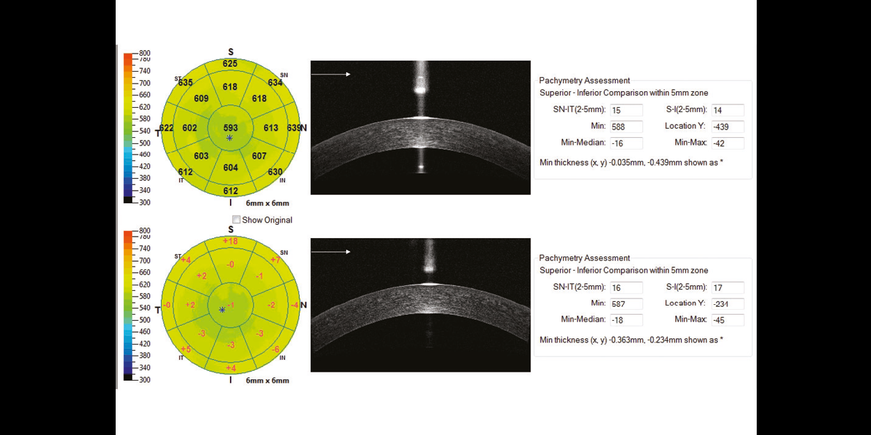
Visualize and quantify 6mm of epithelial, stromal and total corneal thickness tonidentify areas of thickening or thinning related to dry eye disease, keratoconus, or previous refractive surgery. The Change Analysis report measures changes in thickness between visits.

Enables precise visualization of the lens-to-eye relationship for specialty lens fitting. Similar to the pachymetry scan, Optovue iVue80+ iCam 12 systems map the space between the scleral lens and the cornea, providing an accurate clearance measurement.

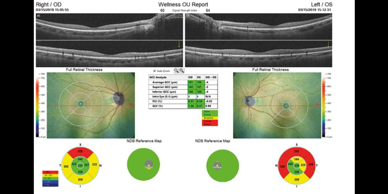
Advanced GCC imaging reveals ganglion cell and axon loss in optic nerve head disease. GCC thickness mapping improves clarity in structural change identification. Optovue’s exclusive FLV% and GLV% analyses increase GCC sensitivity and specificity.

The iWellnessExam is an Optovue exclusive that delivers a quick, easy OCT scan to promote better overall patient eye health. Its usefulness stems from a single, comprehensive report that depicts: retinal thickness and GCC® thickness with normative comparison, symmetry analysis, FLV% and GLV%, proprietary Optovue GCC metrics that provide important information to aid in ocular disease diagnosis and management and eight high-resolution B-scans.

Retina mapping with normative comparison, retina change analysis and 3D retinal imaging with en face presentation are all depicted in high-resolution, easy-to-interpret colour reports, combined with color Fundus photography.

Advanced capabilities include RNFL and GCC® combination reports with normative comparison as well as RNFL and GCC trend analysis — both standard.
Anterior segment capabilities include highly detailed reports for pachymetry mapping, anterior segment angle measurement and Vault Mapping for specialty lens fitting.

3D en face view provides multi-layer, high-resolution virtual dissection of the retina and optic disc, and depicts them in a way that preserves the retina’s natural curvature. This reduces distortion for simpler interpretation and enhanced 3D visual assessment.

High-performance fundus camera that delivers posterior and anterior segment images with exceptional depth, 45° colour and red-free imaging, with image sharpening. The system software is very easy to learn and enables fast, flexible image review right out of the gate. Optovue iVue80+ iCam 12, automatically overlays iVue OCT images onto the fundus photos.

The system software is very intuitive with helpful graphics and timely prompts that walk you through an exam. Most users are up to speed quickly.

Scanner OCT Image : 80,000 A-scan/second
Depth Resolution (in tissue) : 5.0μm
Traverse Resolution : 15μm (retina)
Scan Range Depth : 2 – 2.3mm (retina)
Scan Beam Wavelength : 840nm (+/-10nm)
OCT Fundus Image (En Face) : FOV 12mm(H) x 9mm(V)
Minimum Pupil Diameter : 2.5mm
External Image (Live IR) FOV : 13mm x 9mm
Table Dimensions (in) : (W) 19.1 x (L) 34.4 x (H) 263-343
Operating System : Windows 7, 8 and 10; 65-bit OS compatible
Processor Speed : 3.0 GHz; Intel Quad Core (desktop); Core 2 (laptop)
Network Bandwidth : 1 Gbps or higher
Computer RAM : 4 GB or higher
Monitor Resolution : 1920 x 1080 at 32-bit
iCam 12 Non- Mydriatic Fundus Camera


Screening for refractive surgery
Raise awareness on the importance of Epithelial Thickness Mapping (ETM) in refractive surgery screening.

Retina protocol
SOLIX delivers pristine images of retinal structures with unprecedented views of the vitreous and choroid, that enable confident diagnosis and management of retinal pathologies.
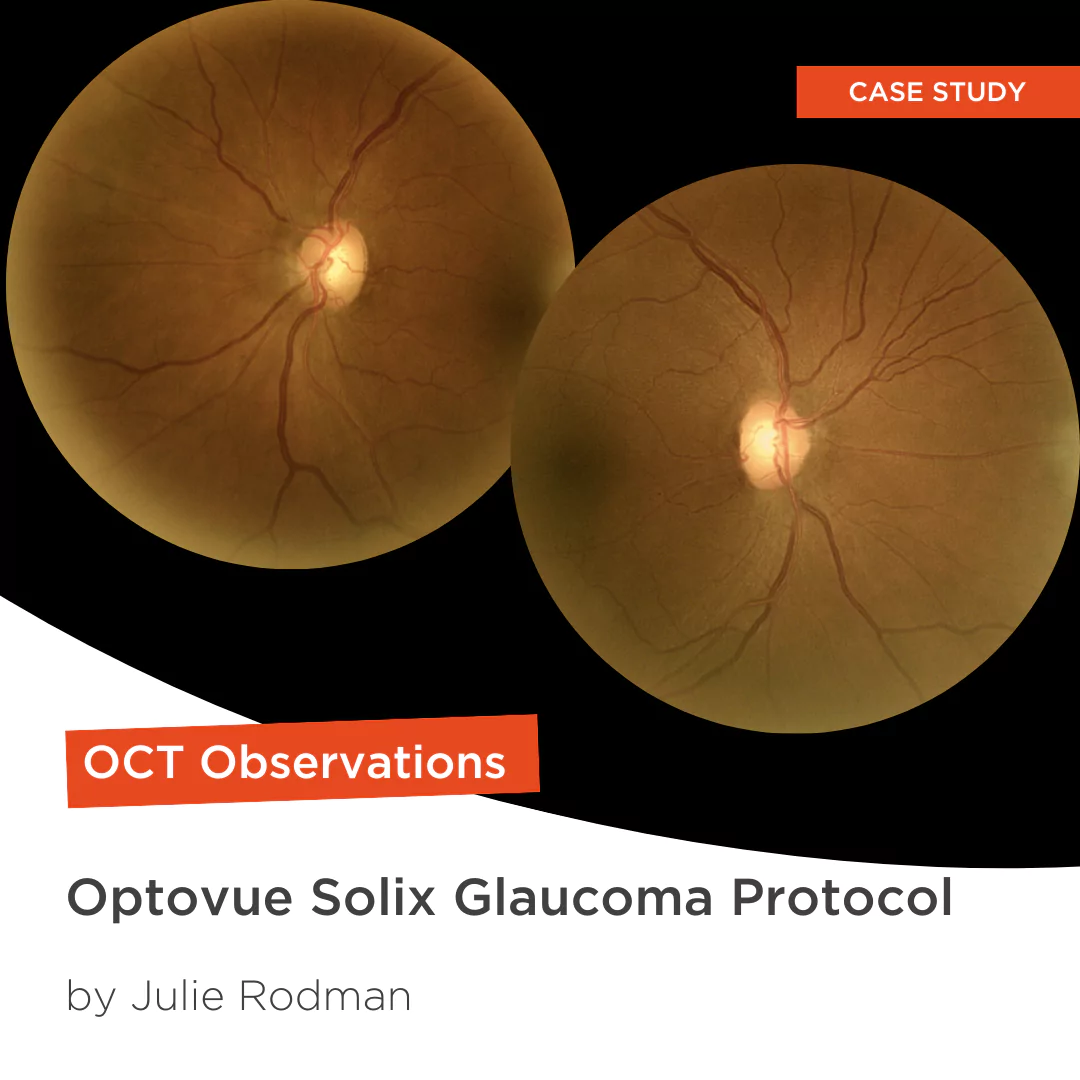
Protocolo Glaucoma
The SOLIX glaucoma package delivers in-depth analysis of the optic nerve head structure and vasculature. Optovue-exclusive scans bring additional insights that aid in clinical decision making.
Protocole Glaucome
The SOLIX glaucoma package delivers in-depth analysis of the optic nerve head structure and vasculature. Optovue-exclusive scans bring additional insights that aid in clinical decision making.
Glaucoma protocol
The SOLIX glaucoma package delivers in-depth analysis of the optic nerve head structure and vasculature. Optovue-exclusive scans bring additional insights that aid in clinical decision making.

We are big fans of Briot
Dr. Gray Sass, OD Wildwood Eyecare
Screening for refractive surgery
Raise awareness on the importance of Epithelial Thickness Mapping (ETM) in refractive surgery screening.
Retina protocol
SOLIX delivers pristine images of retinal structures with unprecedented views of the vitreous and choroid, that enable confident diagnosis and management of retinal pathologies.
Protocolo Glaucoma
The SOLIX glaucoma package delivers in-depth analysis of the optic nerve head structure and vasculature. Optovue-exclusive scans bring additional insights that aid in clinical decision making.
Protocole Glaucome
The SOLIX glaucoma package delivers in-depth analysis of the optic nerve head structure and vasculature. Optovue-exclusive scans bring additional insights that aid in clinical decision making.
Glaucoma protocol
The SOLIX glaucoma package delivers in-depth analysis of the optic nerve head structure and vasculature. Optovue-exclusive scans bring additional insights that aid in clinical decision making.
We are big fans of Briot
Dr. Gray Sass, OD Wildwood Eyecare

My office has had a Briot edger for over 10 years
Dr. Albert Morier Consumer Optical

”Implementing the Briot® Attitude 2 has been effortless. It saves me time so I don’t need to spend extra hours in the office.“
Dr. Douglas King Owner, Family Eye Care Optometry Center

Dr. Paul Karpecki Kentucky Eye Institute
Dr. Paul Karpecki Kentucky Eye Institute

Dr. John Gelles is the director of the specialty contact lens division at The Cornea and Laser Eye Institute – Hersh Vision Group and The CLEI Center for Keratoconus.
Dr. John Gelles is the director of the specialty contact lens division at The Cornea and Laser Eye Institute – Hersh Vision Group and The CLEI Center for Keratoconus.

“I can safely and efficiently practice while also giving the patient confidence they are receiving top notch and modern care.”
Jordan Jones, O.D. Director of Eyecare, Wear Eyewear