


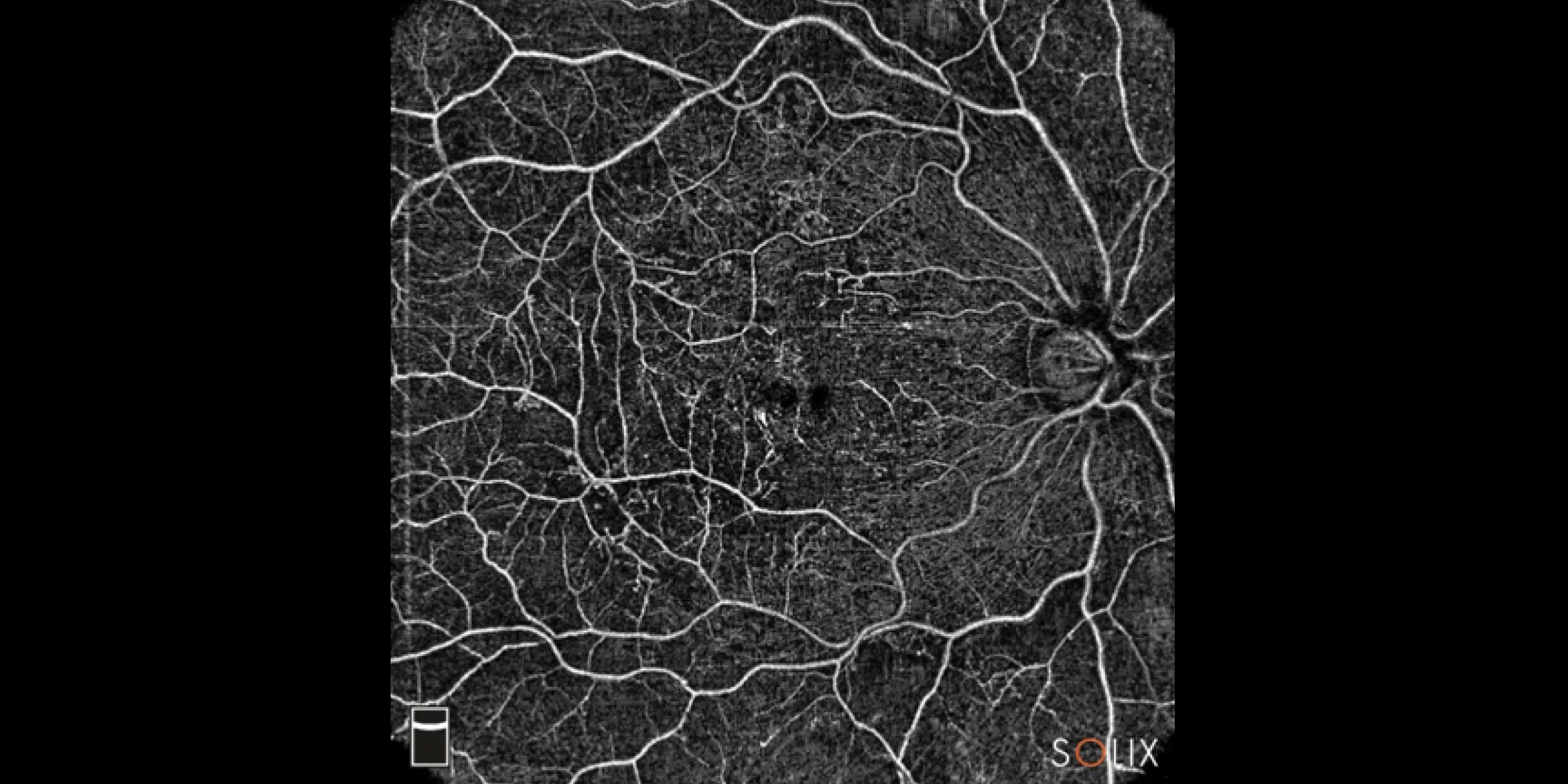
New advanced High-density (HD) retina scan patterns for maximum resolution and post-processing alignment. Tracked High-density scans with SSADA & MCT with vessel-to-vessel post-processing alignment that produces a superior platform for change as it minimizes scan location and movement effects during acquisition and allows for precise registration.

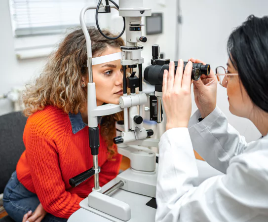
Optovue Solix Essential takes glaucoma scanning to the next level incorporating DualTrac, SSADA, MCT, and AI segmentation along with new features to make an advanced glaucoma system.

Thorough anterior evaluation of pathologies such as keratoconus and dry eye symptoms utilizing 10mm Corneal Layer Map that shows epithelial, stromal, and total corneal thickness with change analysis.

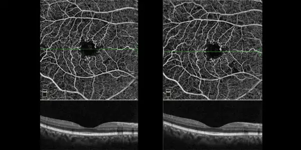
iWellness capabilities have become part of a standard of care for patients suspected of retinal pathologies and/or glaucoma. The Solix AngioWellness scan enables a comprehensive assessment of your diabetic patients and glaucoma suspects by combining structural information on retinal and ganglion cell thickness with objective metrics on retinal vasculature. Utilize FAZ Analytics to uncover early indicators of diabetic changes.
Featuring:

SOLIX ESSENTIAL TECHNICAL SPECIFICATIONS
OCT Imaging | Retina
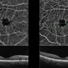
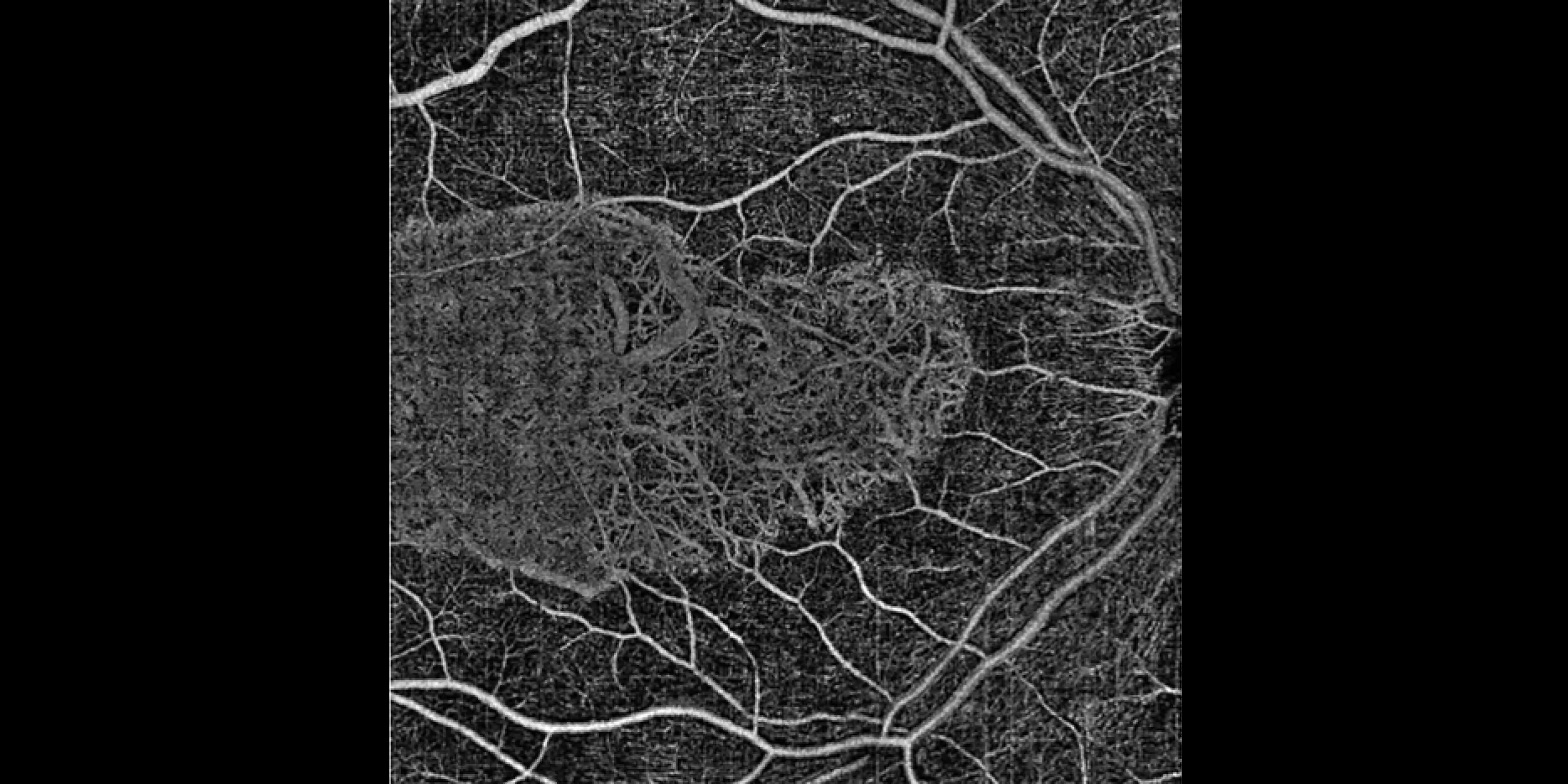
OCT-A Imaging
OCT Imaging | Anterior Segment
Electrical and Physical Specifications
Computer/Networking Specifications


Screening for refractive surgery
Raise awareness on the importance of Epithelial Thickness Mapping (ETM) in refractive surgery screening.

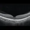
Retina protocol
SOLIX delivers pristine images of retinal structures with unprecedented views of the vitreous and choroid, that enable confident diagnosis and management of retinal pathologies.
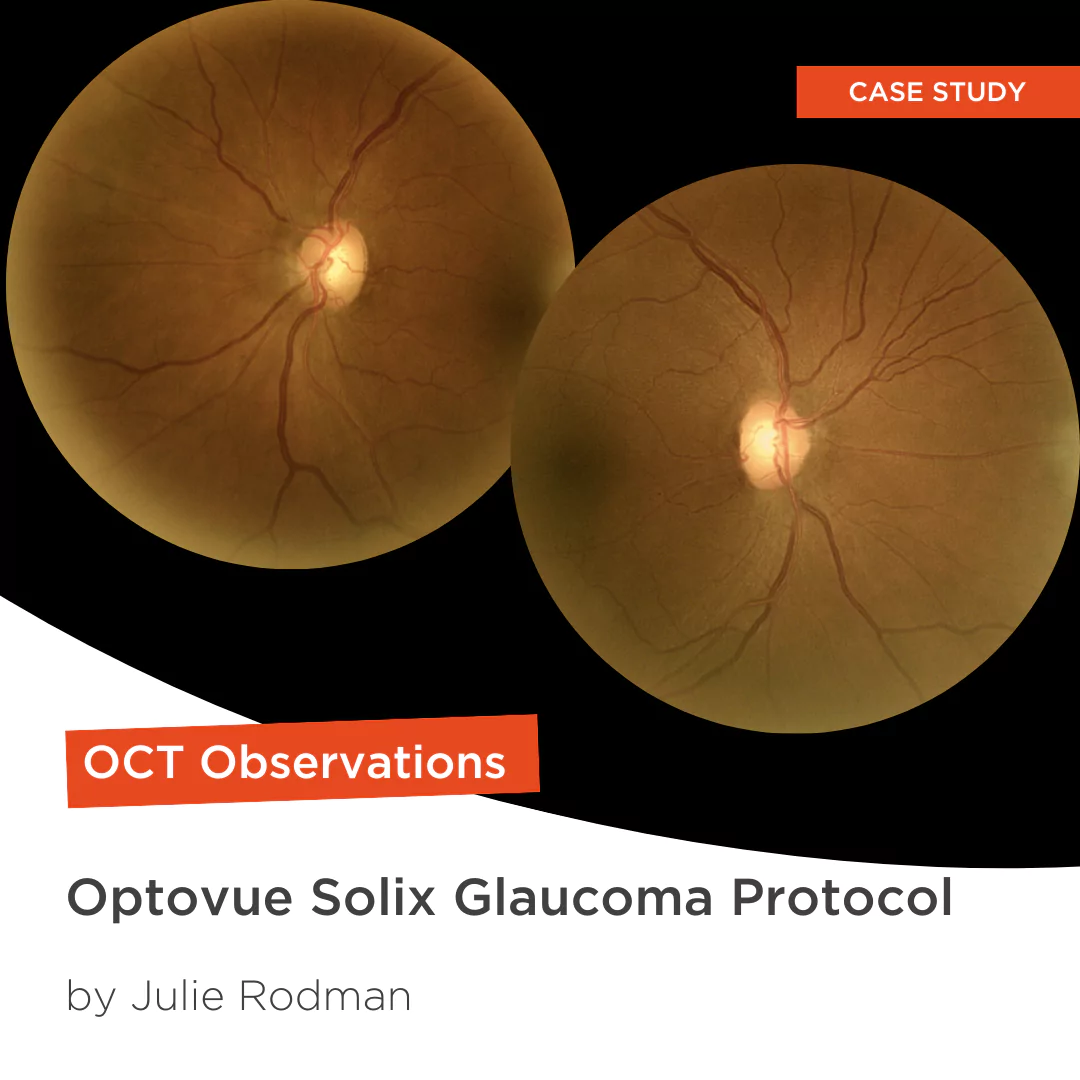
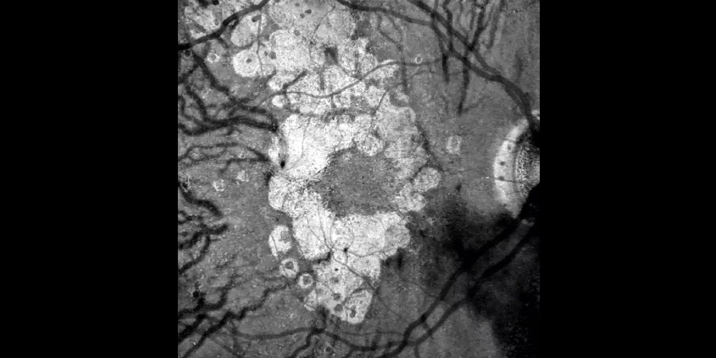
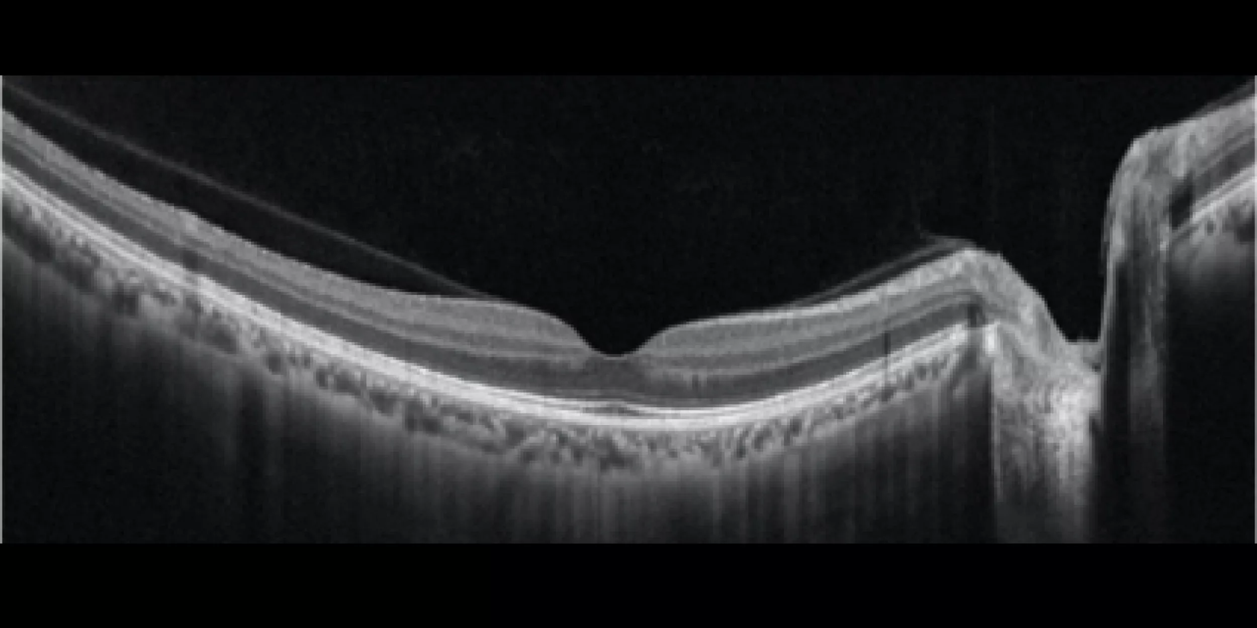
Glaucoma protocol
The SOLIX glaucoma package delivers in-depth analysis of the optic nerve head structure and vasculature. Optovue-exclusive scans bring additional insights that aid in clinical decision making.

We are big fans of Briot
Dr. Gray Sass, OD Wildwood Eyecare

”Implementing the Briot® Attitude 2 has been effortless. It saves me time so I don’t need to spend extra hours in the office.“
Dr. Douglas King Owner, Family Eye Care Optometry Center

My office has had a Briot edger for over 10 years
Dr. Albert Morier Consumer Optical