Visionix VX 650

Bring the retina into focus.

More data. Better visual outcomes.

Provide great vision today, help protect it for a lifetime.

The most comprehensive multimodal diagnostic tool available on the market today.

Diabetic Retinopathy Evaluation

The VX 650 fundus camera helps you to detect and document diabetic retinopathy including vascular abnormalities, hemorrhages, and neovascularization that allows eye care providers to ensure patients receive the care they need before permanent vision loss occurs.

Complete Glaucoma Assessment

VX 650 integrates the fundus camera, scheimpflug camera, tonometer and pachymeter to provide data such as intra-ocular pressure, corneal thickness, irido-corneal angles, and cup-to-disc ratio to fully assess for early changes and identify patients with possible glaucoma.

Retinal Pathology Screening

Most retinal pathologies are not associated with pain and do not have visual symptoms in the initial stages. The VX 650 screens for potentially dangerous retinal findings, such as choroidal nevi, hypertensive retinopathy and drug toxicity.

Early detection of eye diseases: Key to Practice Growth

Accurate Comprehensive 5-minute Exam

This fully automated multi-modal solution allows for the early detection of major ocular pathologies in a single device.  

With precise measurements and analysis, the VX 650 can detect all major pathologies that cause visual impairment or blindness.

Objective day and night refraction using Shack-Hartmann wavefront technology that includes lower-order and higher-order aberrations.

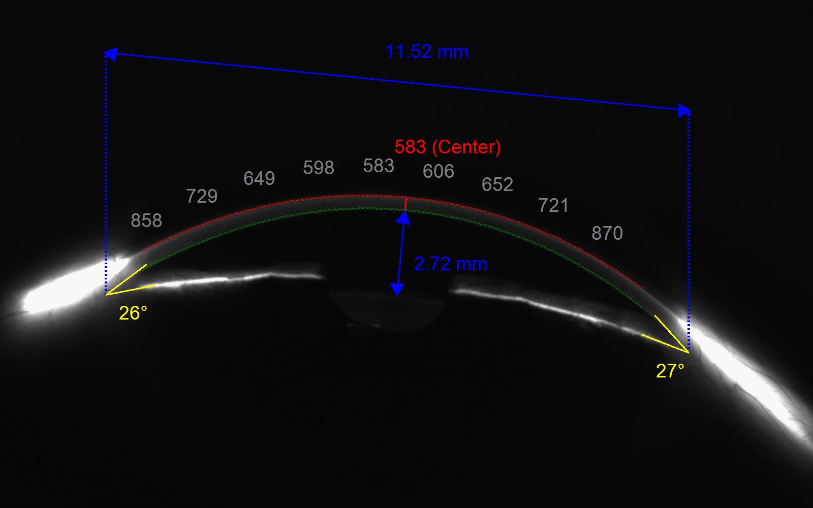
A Scheimpflug camera with retro-illumination capacity visualizes the anterior angle and detects corneal or lens opacities.

Click on the photo to see it in full size.

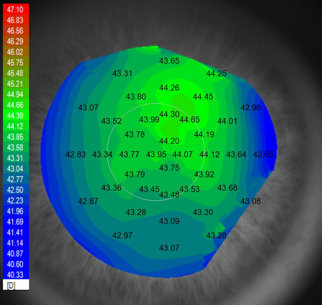
Placido rings measure corneal curvature and maps the corneal surface to detect any irregularities that may indicate pathology.

Click on the photo to see it in full size.

The non-contact tonometer provides a non-invasive measurement of the intra-ocular pressure (IOP)

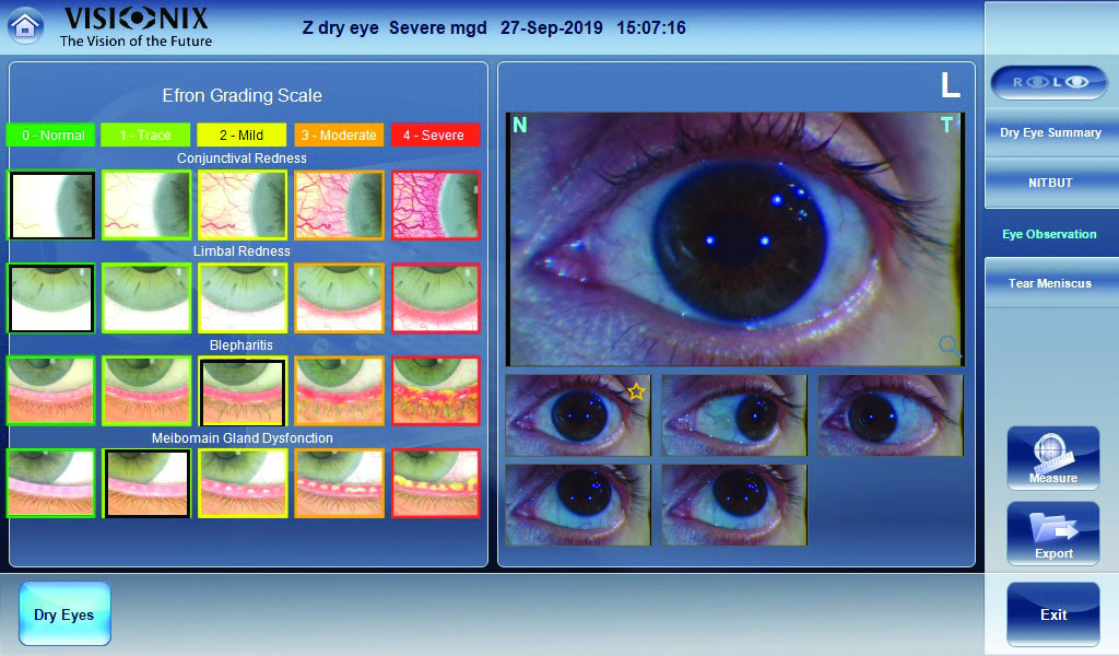
An external camera detects and documents irregularities on the anterior eye including the eyelids, sclera, conjunctiva cornea, iris or pupil.

Click on the photo to see it in full size.

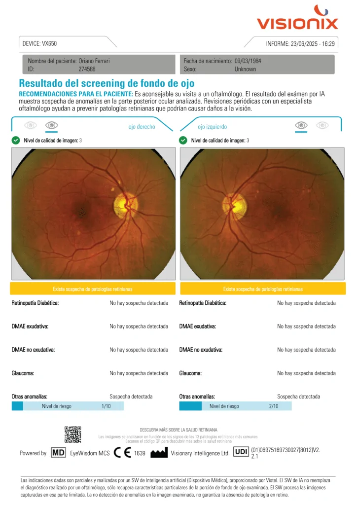
A retinal camera detects retinal changes and allows a throrough review of the fundus to identify retinal pathologies such as DR, ARMD and GLC.

Click on the photo to see it in full size.


The pachymeter measures the central corneal thickness that can indicate certain dystrophies and is used to interpret the IOP.

Click on the photo to see it in full size.

A non-invasive tear break-up time evaluates ocular surface lubrication and is used to assess for dry eye disease.

Click on the photo to see it in full size

Click on the photo to see it in full size.

Better patient flow, less time

Reduces overall patient movement and chair time.

Secure data management

Results are GDPR & HIPAA compliant for data sharing.

Telehealth and remote ready

Can be fully operated remotely and data can be reviewed from anywhere.