


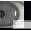

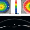
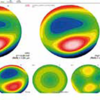
An effective, clinically validated keratoconus screening system provides guidance on ectasia risk, highlighting cases where the likelihood of complications is higher.
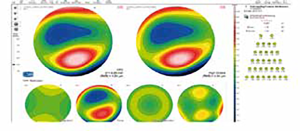
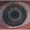
Aberrometric analysis provides a comprehensive overview of corneal aberrations. It is possible to select the anterior, posterior, or total corneal contribution for different pupil diameters. The OPD/WFE map and visual simulations (PSF, MTF, optotype) can assist the clinician in understanding or explaining the patient’s visual discomfort.
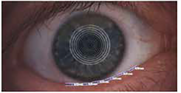
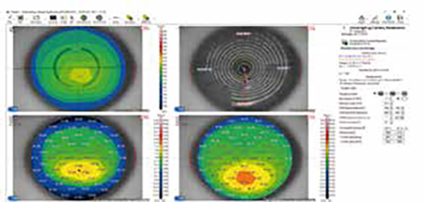
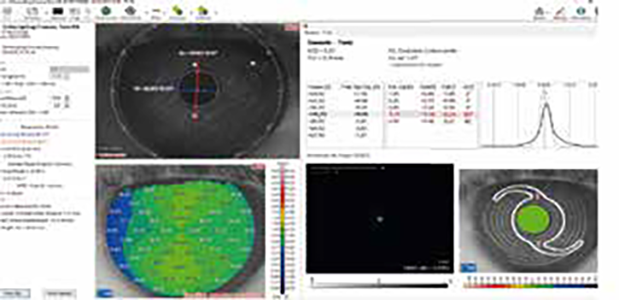
Based on the pachymetric map and corneal elevation data, VX210 enables the planning of intrastromal corneal ring implantation, which may represent a surgical option for the correction of refractive errors and certain forms of keratoconus.
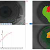
Fully integrated with anterior corneal surface topography, it measures pupil size under scotopic, mesopic, photopic, and dynamic conditions. Knowledge of the pupil center and diameter is essential for all clinical procedures aimed at optimizing visual quality.
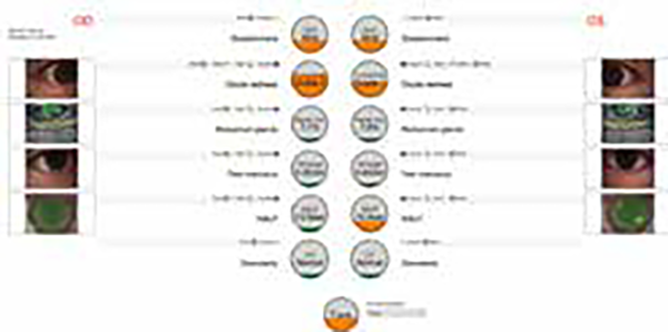
The device is equipped with a white light source for capturing color images or videos, a diffuser filter for analyzing the tear lipid layer, and a cobalt blue light source for assessing rigid contact lens clearance with fluorescein. The magnification adjustment is also particularly useful for capturing images of the tear meniscus and ocular redness
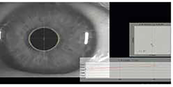
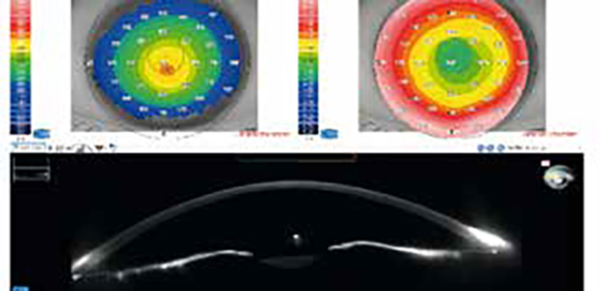
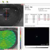
For glaucoma specialists, the device provides measurements of the iridocorneal angles and pachymetry. These two values, used in the most common IOP correction formulas, help in diagnosing the condition when it is related to the configuration of the anterior chamber.


Screening for refractive surgery
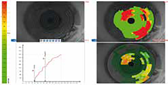
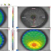
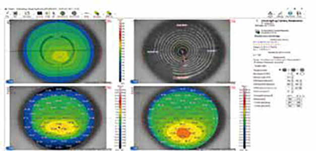
Raise awareness on the importance of Epithelial Thickness Mapping (ETM) in refractive surgery screening.

Retina protocol
SOLIX delivers pristine images of retinal structures with unprecedented views of the vitreous and choroid, that enable confident diagnosis and management of retinal pathologies.
Protocolo Glaucoma
The SOLIX glaucoma package delivers in-depth analysis of the optic nerve head structure and vasculature. Optovue-exclusive scans bring additional insights that aid in clinical decision making.
Protocole Glaucome
The SOLIX glaucoma package delivers in-depth analysis of the optic nerve head structure and vasculature. Optovue-exclusive scans bring additional insights that aid in clinical decision making.
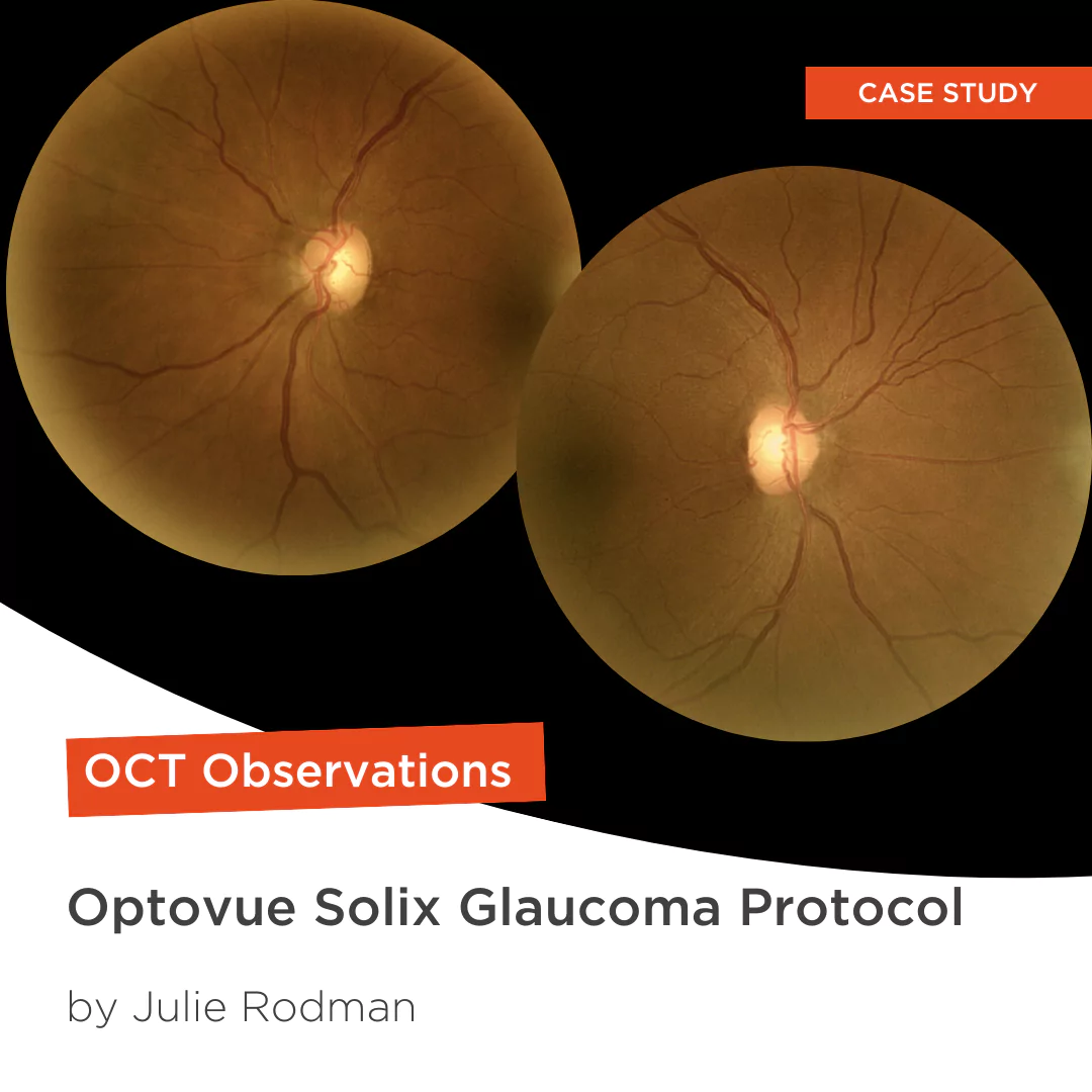
Glaucoma protocol
The SOLIX glaucoma package delivers in-depth analysis of the optic nerve head structure and vasculature. Optovue-exclusive scans bring additional insights that aid in clinical decision making.

We are big fans of Briot
Dr. Gray Sass, OD Wildwood Eyecare
Screening for refractive surgery
Raise awareness on the importance of Epithelial Thickness Mapping (ETM) in refractive surgery screening.
Retina protocol
SOLIX delivers pristine images of retinal structures with unprecedented views of the vitreous and choroid, that enable confident diagnosis and management of retinal pathologies.
Protocolo Glaucoma
The SOLIX glaucoma package delivers in-depth analysis of the optic nerve head structure and vasculature. Optovue-exclusive scans bring additional insights that aid in clinical decision making.
Protocole Glaucome
The SOLIX glaucoma package delivers in-depth analysis of the optic nerve head structure and vasculature. Optovue-exclusive scans bring additional insights that aid in clinical decision making.
Glaucoma protocol
The SOLIX glaucoma package delivers in-depth analysis of the optic nerve head structure and vasculature. Optovue-exclusive scans bring additional insights that aid in clinical decision making.
We are big fans of Briot
Dr. Gray Sass, OD Wildwood Eyecare

My office has had a Briot edger for over 10 years
Dr. Albert Morier Consumer Optical

”Implementing the Briot® Attitude 2 has been effortless. It saves me time so I don’t need to spend extra hours in the office.“
Dr. Douglas King Owner, Family Eye Care Optometry Center

Dr. Paul Karpecki Kentucky Eye Institute
Dr. Paul Karpecki Kentucky Eye Institute

Dr. John Gelles is the director of the specialty contact lens division at The Cornea and Laser Eye Institute – Hersh Vision Group and The CLEI Center for Keratoconus.
Dr. John Gelles is the director of the specialty contact lens division at The Cornea and Laser Eye Institute – Hersh Vision Group and The CLEI Center for Keratoconus.

“I can safely and efficiently practice while also giving the patient confidence they are receiving top notch and modern care.”
Jordan Jones, O.D. Director of Eyecare, Wear Eyewear