A comprehensive multi-modal device for anterior / posterior segments measurement and analysis (ARK, Aberrometer, Topographer, Pachymeter, Scheimpflug camera, Tonometer & now integrating the Fundus camera).

From cornea to retina, it detects all major defects and pathologies, like keratoconus, cataract, glaucoma, nevus, diabetic retinopathy, retinal hemorrhage and more.

Reduce overall patient movement and time in the pre-test room while providing a comprehensive examination to every patient.

Accurate and reproducible results regardless of the operator.

Results available for GDPR (General Data Protection Regulation) and HIPAA (Health Insurance Portability and Accountability Act) compliant data sharing, for either local or remote review.

The device can be fully operated remotely. Additionally, data is available for review by a licensed practitioner – from anywhere.

| Feature | Value |
|---|---|
| Method | Static horizontal scan with the Scheimpflug camera |
| Pachymeter measuring range | 150µm – 1300µm |
| Pachymetry accuracy | < 5µm |
| IC angle measuring range | 0°-60° |
| IC resolution | 0.1° |
| Pupil illumination Blue light | 455nm |
| Feature | Value |
|---|---|
| Number of rings | 24 |
| Number of measuring points | 6.144 |
| Number of points analyzed | More than 100,000 |
| Diameter of covered corneal area at 43D | From 0.75 mm to more than 10 mm |
| Measurement range | From 37.5 D to 56 D |
| Repeatability | 0.03mm |
| Method | Placido rings |
| Feature | Value |
|---|---|
| Measurement range | Calibrated range 7 – 44 mmHg |
| Feature | Value |
|---|---|
| Alignment | XYZ automatic |
| Display | 15.6” . TFT screen Multi-touch screen |
| Observation area | ø 14 mm |
| Medical device directive | EC MDD 93/42/EC modified by directive 2007/47/EC |
| Output | RS232 / USB / VGA / LAN / HDMI / DP |
| Feature | Value |
|---|---|
| Spherical power range | -20D to +20D |
| Cylinder power range | 0D to ±8D |
| Axis | 0 to 180° |
| Measuring area | Min. ø 2mm – Max. 7mm (3zones) |
| Number of measuring points | 6.144 points for 7mm pupil at 0D |
| Acquisition time | 0.2sec |
| Method | Shack-Hartmann |
| Feature | Value |
|---|---|
| Angle of view | 45° |
| Resolution | 6Mpix |
| Optical resolution | > 60 lines/mm |


Screening for refractive surgery
Raise awareness on the importance of Epithelial Thickness Mapping (ETM) in refractive surgery screening.

Retina protocol
SOLIX delivers pristine images of retinal structures with unprecedented views of the vitreous and choroid, that enable confident diagnosis and management of retinal pathologies.
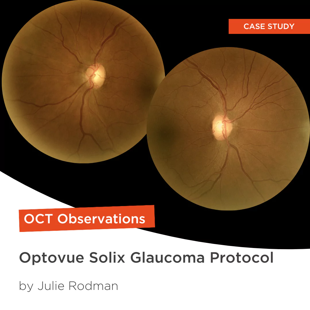
Glaucoma protocol
The SOLIX glaucoma package delivers in-depth analysis of the optic nerve head structure and vasculature. Optovue-exclusive scans bring additional insights that aid in clinical decision making.

We are big fans of Briot
Dr. Gray Sass, OD Wildwood Eyecare

”Implementing the Briot® Attitude 2 has been effortless. It saves me time so I don’t need to spend extra hours in the office.“
Dr. Douglas King Owner, Family Eye Care Optometry Center

My office has had a Briot edger for over 10 years
Dr. Albert Morier Consumer Optical