


| Feature | Value |
|---|---|
| AR-K | • |
| Occular Aberro. | • |
| Retro | • |
| Corneal Topograph | • |
| Non Contact Tonometer | • |
| Feature | Value |
|---|---|
| Pachymetry | • |
| Full Eye Tracking | • |
| Back surface of the cornea | |
| Remote Acces | • |
| Offline/Webservice | • |
| Colour camera |
| Feature | Value |
|---|---|
| NBUT | |
| Efron classification | |
| Tear meniscus value |
| Feature | Value |
|---|---|
| Alignment | XYZ automatic |
| Display | 10.1” (1 024 x 600) TFT multi-touch screen |
| Observation area | ø 14 mm |
| Medical device directive | EC MDD 93/42/EC modified by directive 2007/47/EC |
| Output | RS232 / USB / VGA / LAN |
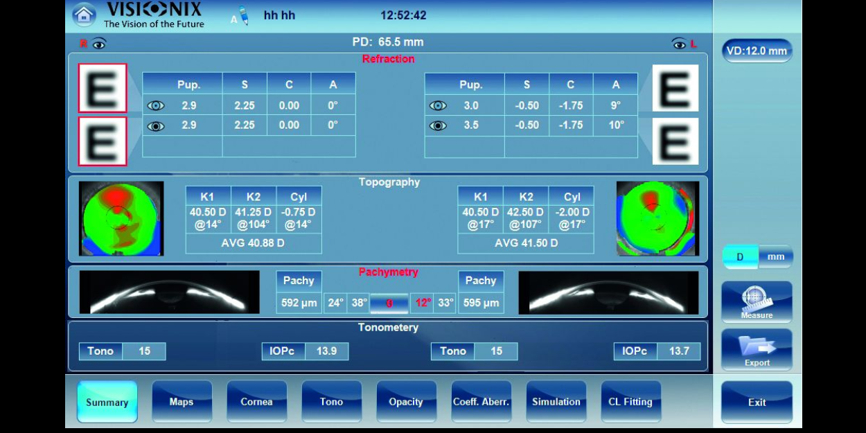
| Feature | Value |
|---|---|
| Spherical power range | -20D to +20D |
| Cylinder power range | 0D to + 8D |
| Axis | 0 to 180° |
| Measuring area | Min. ø 2 mm – Max. 7 mm (3 zones) |
| Number of measuring points | 1,300 points |
| Acquisition time | 0.2 sec |
| Method | Shack-Hartmann |
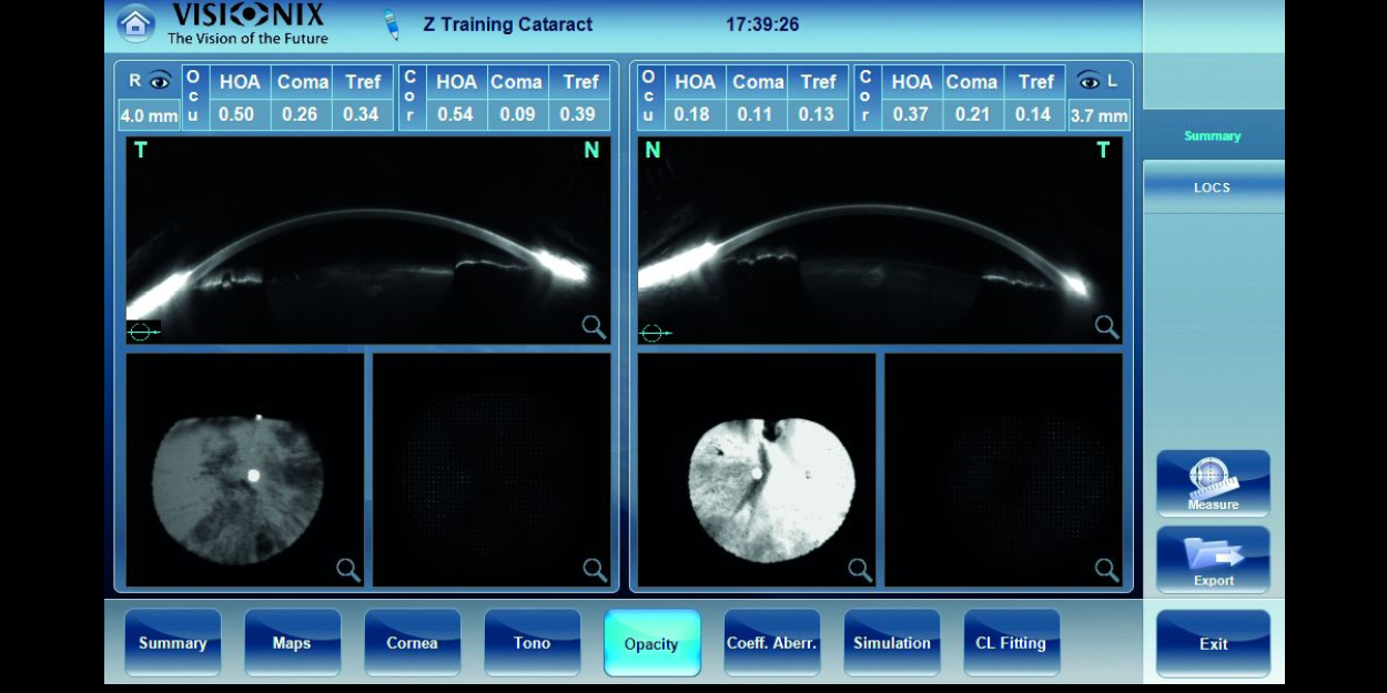
| Feature | Value |
|---|---|
| Method | Continuous horizontal scan with the Scheimpflug camera |
| Pachymeter measuring range | 150-1300 μm |
| Pachymeter resolution | +/- 10 microns |
| IC angle measuring range | 0°-60° |
| IC resolution | 0.1° |
| Pupil illumination | Blue light 455 nm |
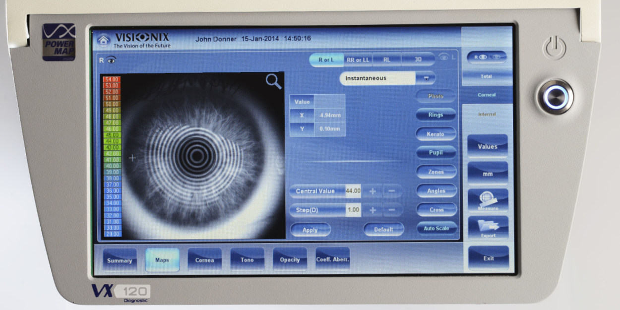
| Feature | Value |
|---|---|
| Number of rings | 24 |
| Number of measuring points | 6.144 |
| Number of points analyzed | More than 100,000 |
| Diameter of covered corneal area at 43D | From 0.75 mm to more than 10 mm |
| Measurement range | From 37.5 D to 56 D |
| Repeatability | 0.02 D |
| Method | Placido rings |
| Feature | Value |
|---|---|
| Measurement range | 7 mmHg to 44 mmHg |


Il a été facile d'intégrer la Briot Attitude 2 dans mon activité. Elle me fait gagner du temps, ce qui m'évite de faire des heures supplémentaires au bureau.
Dr. Douglas King Owner, Family Eye Care Optometry Center