


| Feature | |
|---|---|
| AR-K | • |
| Occular Aberro. | • |
| Retro | • |
| Corneal Topographer | • |
| Feature | |
|---|---|
| Pachymetry | • |
| Full Eye Tracking | • |
| Back surface of the cornea | |
| Remote Acces | • |
| Offline/Webservice | • |
| Feature | |
|---|---|
| Alignment | XYZ automatic |
| Display | 10.1” (1 024 x 600) TFT multi-touch screen |
| Observation area | ø 14 mm |
| Medical device directive | EC MDD 93/42/EC modified by directive 2007/47/EC |
| Output | RS232 / USB / VGA / LAN |
| Feature | |
|---|---|
| Spherical power range | -20D to +20D |
| Cylinder power range | 0D to + 8D |
| Axis | 0 to 180° |
| Measuring area | Min. ø 2 mm – Max. 7 mm (3 zones) |
| Number of measuring points | 1,300 points |
| Acquisition time | 0.2 sec |
| Method | Shack-Hartmann |
| Feature | |
|---|---|
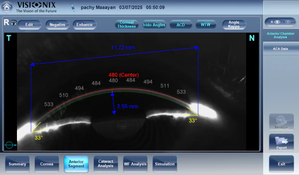
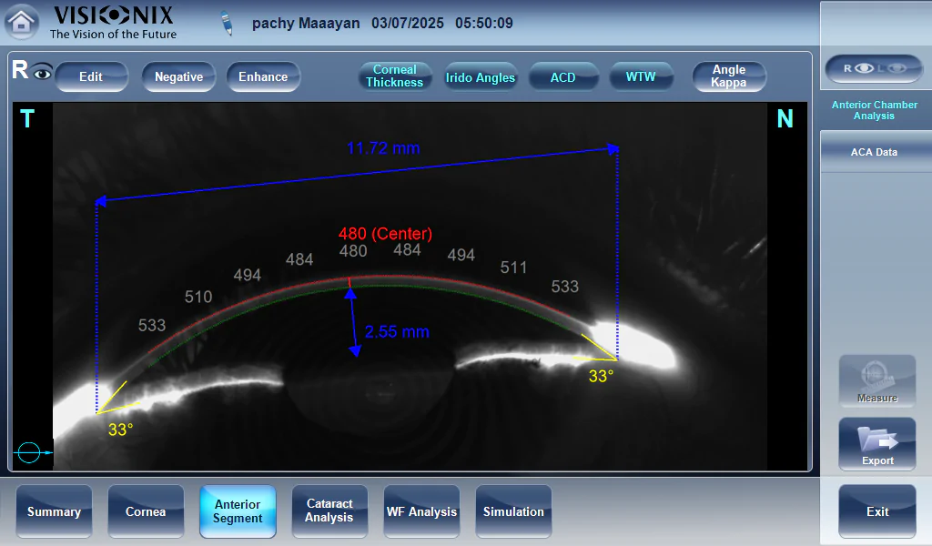
| Method | Continuous horizontal scan with the Scheimpflug camera |
| Pachymeter measuring range | 150-1300 μm |
| Pachymeter resolution | +/- 10 microns |
| IC angle measuring range | 0°-60° |
| IC resolution | 0.1° |
| Pupil illumination | Blue light 455 nm |
| Feature | |
|---|---|
| Number of rings | 24 |
| Number of measuring points | 6.144 |
| Number of points analyzed | More than 100,000 |
| Diameter of covered corneal area at 43D | From 0.75 mm to more than 10 mm |
| Measurement range | From 37.5 D to 56 D |
| Repeatability | 0.02 D |
| Method | Placido rings |


Screening for refractive surgery
Raise awareness on the importance of Epithelial Thickness Mapping (ETM) in refractive surgery screening.

Retina protocol
SOLIX delivers pristine images of retinal structures with unprecedented views of the vitreous and choroid, that enable confident diagnosis and management of retinal pathologies.
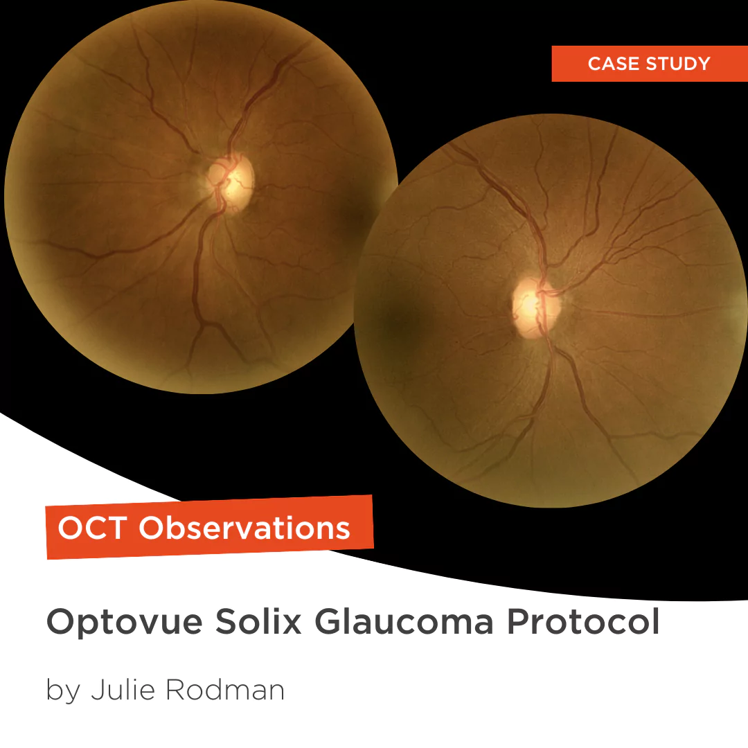
Glaucoma protocol
The SOLIX glaucoma package delivers in-depth analysis of the optic nerve head structure and vasculature. Optovue-exclusive scans bring additional insights that aid in clinical decision making.

We are big fans of Briot
Dr. Gray Sass, OD Wildwood Eyecare

”Implementing the Briot® Attitude 2 has been effortless. It saves me time so I don’t need to spend extra hours in the office.“
Dr. Douglas King Owner, Family Eye Care Optometry Center

My office has had a Briot edger for over 10 years
Dr. Albert Morier Consumer Optical