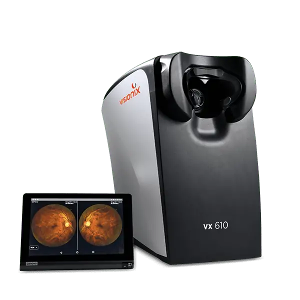


The VX 610 camera utilizes innovative cross-polarized light technology, which provides excellent image quality while maintaining a compact footprint.

The VX 610 is a non-mydriatic retinal camera that allows the observation, capture, and recording of high-resolution images without the need for dilating the pupil.

The VX 610 is designed to be incredibly easy to operate and requires minimal personnel training.

The intuitive interface facilitates a quick start and smooth navigation.

A small footprint and ergonomic design ensure the VX 610 will fit within your practice layout.

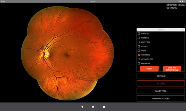
Field of view : 45°
Field of view with mosaic function : 90°
Retinal resolution : 14 microns
Fixations : 7 fixations set-ups
Tablet dimensions : 10.4’’
Weight : 32,4lbs
Size : 13.4 x 16.9 x 18.1 in.
Image filters (vascular, choroidal, nerve fibre, red free)
Cup-to-disc ratio for glaucoma screening
Automatic mosaic function
Shared intranet folder
DICOM standard compatible
Fundus adapter to transfer images to a local PC
Cloud-based secure storage
Client-side data encryption
Automatic database backup
Review images from the password protected website, a network shared folder or a local PC
Multilevel account
Access by personal user


Screening for refractive surgery
Raise awareness on the importance of Epithelial Thickness Mapping (ETM) in refractive surgery screening.

Retina protocol
SOLIX delivers pristine images of retinal structures with unprecedented views of the vitreous and choroid, that enable confident diagnosis and management of retinal pathologies.
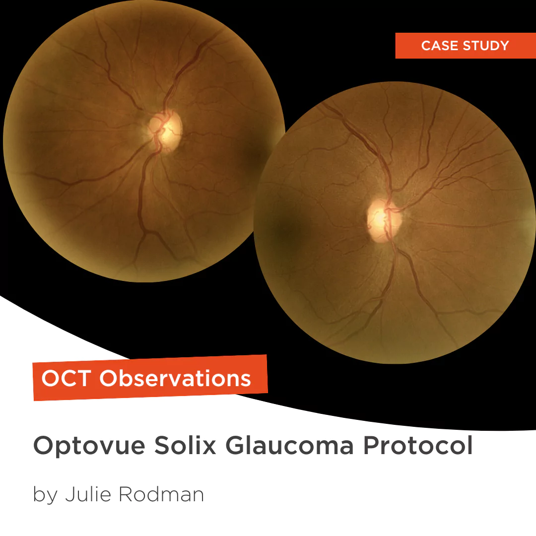
Glaucoma protocol
The SOLIX glaucoma package delivers in-depth analysis of the optic nerve head structure and vasculature. Optovue-exclusive scans bring additional insights that aid in clinical decision making.

We are big fans of Briot
Dr. Gray Sass, OD Wildwood Eyecare

”Implementing the Briot® Attitude 2 has been effortless. It saves me time so I don’t need to spend extra hours in the office.“
Dr. Douglas King Owner, Family Eye Care Optometry Center

My office has had a Briot edger for over 10 years
Dr. Albert Morier Consumer Optical